


Asperoteuthis acanthoderma
Richard E. Young and Clyde F. E. Roper
Introduction
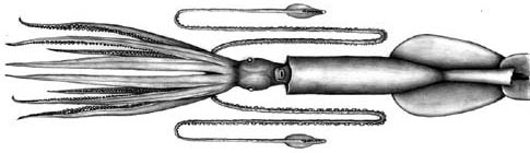
A. acanthoderma reaches a rather large size. The largest specimen known has a mantle length of 78 cm and long, slender tentacles. In one squid (45 cm ML) the tentacles were over 12 times longer than the mantle (i.e., about 5.5 m)(Tsuchiya and Okutani, 1993). The most distinctive feature of this species is the presence of very small, pointed cartilagenous tubercules over the surface of the head, mantle and arms.
Brief diagnosis
An Asperoteuthis ...
- with tiny, pointed tubercules in skin.
- with a Y-shaped groove in the funnel locking-apparatus.
Characteristics
- Arms
- Large arm suckers with 3-4 broadly rounded teeth on distal half of inner ring.
Click on an image to view larger version & data in a new window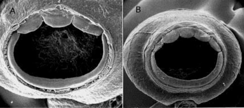
Figure. Oral view of arm suckers of A. acanthoderma: A - Sucker no. 11 counting from arm base, from arm III of the holotype, 190 mm ML. B - Arm sucker from (((WH354))). Scanning electron migrographs by R. Young.
- Tentacles
- Suckers with nine blunt teeth in contact at base over distal half of inner ring.
Click on an image to view larger version & data in a new window
Figure. Tentacular club of A. acanthoderma. Left - Oral view of club sucker from (((WH354))). Scanning electron micrograph by R. Young. Right - Oral view of club. Illustration by J. R. Schroeder.
- Integument
- Covered (except for tentacles) with tiny, conical, cartilagenous tubercules.
Click on an image to view larger version & data in a new window
Figure. A - Cross-section through the mantle of A. acanthoderma. Note the highly vacuolated mantle tissue, the two large, elongate "holes" in the integument which house chromatophores and the conical tubercule (arrow) on the surface of the skin. B - Scanning electron micrograph of mantle surface. Arrows indicate position of tubercules. Photographs from Lu, 1977, with permission.
- Funnel
- Funnel-locking apparatus with long, slender tragus and narrow antitragus that form an inverted Y-shaped groove.
Click on an image to view larger version & data in a new window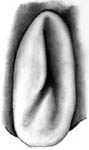
Figure. Frontal view of the funnel component of the funnel/mantle locking apparatus of A. acanthoderma. Drawing by J. R. Schroeder.
- Funnel-locking apparatus with long, slender tragus and narrow antitragus that form an inverted Y-shaped groove.
- Fins
- Fin length 50-55% of ML.
- Fin width 11-13% of ML
- Secondary "fin" comparable in length to primary (= true) fin but more slender.
- Photophores
- Oval photophore on the ventral surface of each eye.
- Numerous photogenic pads on the tentacle stalk.
- Large club-tip photophore with short, broad, terminal papilla.
- Twelve pairs of small, embedded photophores on aboral surface of club in two series and of two sizes.
Click on an image to view larger version & data in a new window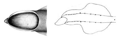
Figure. Aboral views of the tentacular club of A. acanthoderma. Left - Aboral view of the terminal photophore on the tentacular club. Drawing by J. R. Schroeder. Right - Aboral view of tentacle club showing the position of embedded photophores at club base and on either side of the club "stalk" , 180 mm ML. Drawing from Lu, 1977, with permission.
- Measurements
- Arms: Arms I ca. 75-85% of ML; Arms III ca.95-110 % of ML; Arms IV ca. 95-115% 0f ML.
- Tentacular club: Club length ca. 16% of ML; Protective membranes - Proximal set with x-xx trabeculae (xx-xx% of club length); distal set with xx trabeculae (xx-xx% of CL).
- Head: Head length 26% of ML.
Life History
A large 78 cm ML specimen described by Tsuchia and Okutani (1993) was an immature female. Mature stages and doratopsis stages are unknown (however, see comments under Asperoteuthis: Life History).
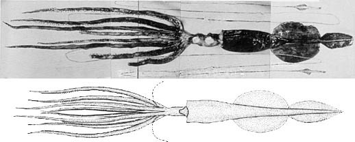
Figure. Ventral views of A. acanthoderma. Top - 560 mm ML, immature female. Note the extremely long tentacles and the dark pigmentation. Bottom - 780 mm ML, immature female. Drawing and photograph from Tsuchiya and Okutani, 1993, with permission.
Distribution
The type locality is the Celebes Sea. This species is also known from waters off Okinawa (Tsuchiya and Okutani, 1993) and Hawaii.
References
Lu, C. C. 1977. A new species of squid Chiroteuthis acanthoderma, from the Southwest Pacific (Cephalopoda, Chiroteuthidae). Steenstrupia, 4: 179-188.
Nesis, K. N. 1980. Taxonomic position of Chiroteuthis famelica Berry. Bull. Moscow Obslich. Ispyt. Prirody, Section Biology, 85: 59-66. [In Russian].
Tsuchiya, K. and T. Okutani (1993). Rare and interesting squids in Japan -X. Recent occurrences of big squids from Okinawa. Venus, 52: 299-311.
Young, R. E. (1991). Chiroteuthid and related paralarvae from Hawaiian waters. Bull. Mar. Sci., 49: 162-185.
Title Illustrations

| Scientific Name | Asperoteuthis acanthoderma |
|---|---|
| Creator | J. R. Schroeder |
| Copyright |
©

|
About This Page
Richard E. Young
University of Hawaii, Honolulu, HI, USA
Clyde F. E. Roper
Smithsonian Institution, Washington, D. C., USA
Page copyright © 1999 Richard E. Young and
- Content changed 08 March 2007
Citing this page:
Young, Richard E. and Roper, Clyde F. E. 2007. Asperoteuthis acanthoderma . Version 08 March 2007 (under construction). http://tolweb.org/Asperoteuthis_acanthoderma/19466/2007.03.08 in The Tree of Life Web Project, http://tolweb.org/


