


Cirrothauma murrayi
Richard E. Young and Michael Vecchione

Introduction
C. murrayi is virtually blind. The eye lacks a lens; the retina is reduced and continuous with a cornea. The eye is embedded within the gelatinous tissue of the head without connection to the surface. Nevertheless, the eye still functions as a simple photoreceptor that cannot form images (Aldred et al, 1983). This species is worldwide in distribution and occupies great depths primarily between 1500 and 4500 m (Nesis, 1982/87) although it has been captured at the surface in an ice hole in the Arctic Ocean (Aldred et al., 1983).
Diagnosis
A Cirrothauma ...
- with reduced eyes lacking lenses.
Characteristics
- Eyes
- Each eye small and without lens.
- Outer eyelid absent. Click on an image to view larger version & data in a new window
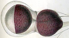
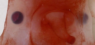
Figure. Left - Eye and white body of C. murrayi. Drawing from Chun, 1910. Right - Ventral view of the eyes, mantle opening and funnel of a fresh C. murrayi. Photograph by M. Vecchione.
- Suckers
- Proximal 6 suckers sessile.
- Suckers over most of arm tiny on long, spindle-shaped, gelatinous stalks.
- Sucker orifice minute or absent.
- Acetabulum absent. Click on an image to view larger version & data in a new window
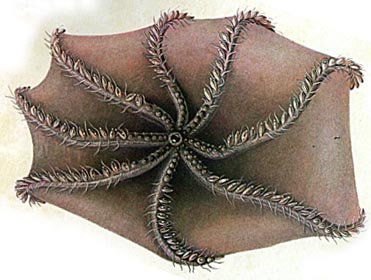
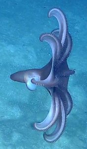
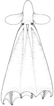
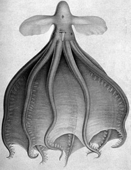
Figure. Oral view of C. murrayi, type illustration, 37 mm ML. Drawing from Chun (1910). Right - Photograph from an ROV of a probable C. murrayi floating near the ocean floor over the Cayman Rise, Carribean Sea. © Ocean Exploration Trust / Sea Research Foundation
Click on an image to view larger version & data in a new window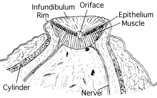 Click on an image to view larger version & data in a new window
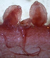
Figure. Left - Longitudinal section through a sucker of C. murrayi, male, 155 mm ML, North Atlantic. Drawing from Aldred et al (1983). The drawing is a section through the tiny sucker that lies at the tip of the long, swollen stalks. The structure labeled "cylinder"appears to consist of acid mucopolysaccharide-containing tissue and internal to it are many small bundles of longitudinal muscle fibres (not shown in drawing) (Aldred et al, 1983). Right - Side view of a portion of an arm of C. murrayi showing the swollen sucker stalks and the tiny apical suckers, fresh (=unfixed) octopod. Photograph by M. Vecchione, R/V G. O. Sars, Mar-Eco cruise, central North Atlantic.
- Web
- Distal web attachments to each arm symmetrical.
- Photophores
- Possible photophore: a small structure located internally at the base of each sucker stalk.
Comments
The above description is taken from Chun (1910) and Aldrich, et al. (1983). More details of the description can be found here.
The reduced suckers are still thought to provide suction but clearly by a somewhat different mechanism than typically seen in octopods. Contraction of the radial muscles near the center of a flat infundibulum that adheres to a surface, should provide suction. Aldred et al. (1983) suggest, however, that adhesion may arise from capillary action of the many minute tubes that make up the cuticle of the infundibulum and that this is especially effective in very deep-living octopods.
Distribution
Type locality: The North Atlantic at 48°24'N, 36°53'W. It is known from the Pacific, Atlantic and Arctic Oceans. In the Northeast Atlantic one study that captured 27 individuals, found they occurred from 2430-4846 m depth, with most captures below 3000 m (Collins, et al., 2001).

Figure. Photograph form an ROV of a probable C. murrayi swimming near the ocean floor over the Cayman Rise, Carribean Sea. © Ocean Exploration Trust / Sea Research Foundation.
References
Aldred, R. G., M. Nixon and J. Z. Young. 1983. Cirrothauma murrayi Chun, a finned octopod. Phil. Trans. Roy. Soc. Lond., 301: 1-54.
Collins, M. A., C. Yau, L. Allcock and M. H. Thurston. 2001. Distribution of deep-water benthic and bentho-pelagic cephalopods from the north-east Atlantic. Jour. Mar. Biol. Ass. U.K., 81: 105-117.
Chun, C. 1910. Die Cephalopoden. Oegopsida. Wissenschaftliche Ergebnisse der Deutschen Tiefsee-Expedition, "Valdivia" 1898-1899, 18: 1-522 + Atlas.
Nesis, K. N. 1982/87. Abridged key to the cephalopod mollusks of the world's ocean. 385+ii pp. Light and Food Industry Publishing House, Moscow. (In Russian.). Translated into English by B. S. Levitov, ed. by L. A. Burgess (1987), Cephalopods of the world. T. F. H. Publications, Neptune City, NJ, 351pp.
Title Illustrations

| Scientific Name | Cirrothauma murrayi |
|---|---|
| Location | Antarctic waters |
| Creator | E. McSweeny |
| Copyright | © 1996 E. McSweeny |

| Scientific Name | Cirrothauma murrayi |
|---|---|
| Location | North Atlantic |
| Reference | Aldred, R. G., M. Nixon and J. Z. Young. 1983. Cirrothauma murrayi Chun, a finned octopod. Phil. Trans. Roy. Soc. Lond., 301: 1-54. |
| Sex | Male |
| Size | 155 mm ML |
| Copyright | © 1983 R.G. Aldred et al. |

| Scientific Name | Cirrothauma murrayi |
|---|---|
| Reference | Chun, C. 1910. Die Cephalopoden. Oegopsida. Wissenschaftliche Ergebnisse der Deutschen Tiefsee-Expedition, "Valdivia" 1898-1899, 18: 1-522 + Atlas. |
| Size | 37 mm ML |
About This Page

University of Hawaii, Honolulu, HI, USA
National Museum of Natural History, Washington, D. C. , USA
Page copyright © 2016 and
 Page: Tree of Life
Cirrothauma murrayi .
Authored by
Richard E. Young and Michael Vecchione.
The TEXT of this page is licensed under the
Creative Commons Attribution-NonCommercial License - Version 3.0. Note that images and other media
featured on this page are each governed by their own license, and they may or may not be available
for reuse. Click on an image or a media link to access the media data window, which provides the
relevant licensing information. For the general terms and conditions of ToL material reuse and
redistribution, please see the Tree of Life Copyright
Policies.
Page: Tree of Life
Cirrothauma murrayi .
Authored by
Richard E. Young and Michael Vecchione.
The TEXT of this page is licensed under the
Creative Commons Attribution-NonCommercial License - Version 3.0. Note that images and other media
featured on this page are each governed by their own license, and they may or may not be available
for reuse. Click on an image or a media link to access the media data window, which provides the
relevant licensing information. For the general terms and conditions of ToL material reuse and
redistribution, please see the Tree of Life Copyright
Policies.
- First online 13 May 2003
- Content changed 27 February 2016
Citing this page:
Young, Richard E. and Michael Vecchione. 2016. Cirrothauma murrayi . Version 27 February 2016 (under construction). http://tolweb.org/Cirrothauma_murrayi/20099/2016.02.27 in The Tree of Life Web Project, http://tolweb.org/


