


Amphilina foliacea
Klaus RohdeIntroduction
The species is widely distributed in Europe and Asia excluding western and southwestern Europe and eastern and southern Asia. Janicki, who worked out the life cycle of the species, suggested that the original life cycle, in the evolutionary past, included a crustacean first and a fish second intermediate host as well as an extinct final host that fed on the fish, harbouring the adult parasite in its digestive tract (Fig. 9). Justification for this hypothesis is that, in other cestodes, stages infecting the body cavity of a fish host are larval forms (plerocercoids). The hypothetical original life cycle would resemble that of Diphyllobothrium latum, which uses crustaceans as first and fish as second intermediate, and fish-eating mammals as final hosts. The maximum length of specimens is 65 mm, the maximum width 30 mm.
The life cycle of this species is described on the Life Cycles page. For information about pathology see the Effects on the Host page.
Characteristics

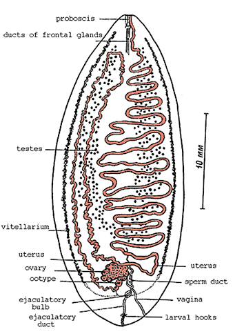
Figure 1. Amphilina foliacea from sturgeon. Note the location of testes between uterine coils, and well separated vaginal and male pores at posterior end of body. According to Dubinina (1962), redrawn from Dubinina (1982).
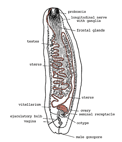
Figure 2. Amphilina foliacea from the body cavity of the intermediate host, Pontogammarus robustoides. Note advanced stage of development of male and female reproductive organs. According to Dubinina (1974), redrawn from Dubinina (1982).
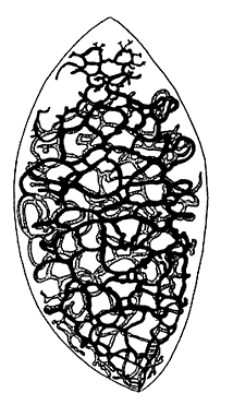
Figure 3. Excretory ducts of Amphilina foliacea. According to Hein (1904), from Dubinina (1982).
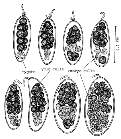
Figure 4. Early cleavage stages of Amphilina foliacea. Note stalk at one pole of egg, dark yolk cells and lighter egg- and cleavage cells. According to Dubinina (1974), from Dubinina (1982).
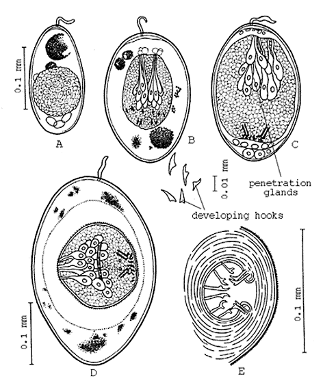
Figure 5. Advanced stages in larval development of Amphilina foliacea. Note change in the shape of the hooks and development of penetration glands. According to Dubinina (1974), from Dubinina (1982).
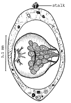
Figure 6. Fully developed larva of Amphilina foliacea in egg. Redrawn from Dubinina (1982).
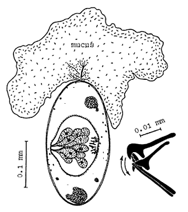
Figure 7. Fully developed larva of Amphilina foliacea with mucus exuded into water through stalk of egg, and larval hooks observed in vivo acting antagonistically in the directions indicated by the arrows. According to Dubinina (1974), from Dubinina (1982).
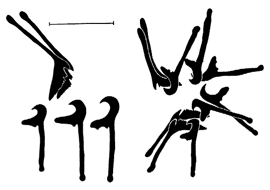
Figure 8. Larval hooks of Amphilina foliacea. Left: one of each pair drawn separately, right: all ten hooks drawn as seen in the larva. Note two pairs of serrate hooks, three pairs of halberd-shaped hooks of different sizes. Scale = 0.02 mm. According to Dubinina (1972), from Dubinina (1982).
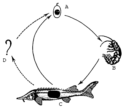
Figure 9. Life cycle of Amphilina foliacea according to Janicki (1930). A egg, B amphipod intermediate host, C sturgeon final host, D hypothetical extinct final host. From Dubinina (1982).
References
Dubinina, M.N. (1982). Parasitic worms of the class Amphilinida (Platyhelminthes). "Nauka", Leningrad (in Russian). (Older references therein).
About This Page
Klaus Rohde

University of New England, Armidale, New South Wales, Australia
Correspondence regarding this page should be directed to Klaus Rohde at
Page copyright © 2000 Klaus Rohde
 Page: Tree of Life
Amphilina foliacea.
Authored by
Klaus Rohde.
The TEXT of this page is licensed under the
Creative Commons Attribution License - Version 3.0. Note that images and other media
featured on this page are each governed by their own license, and they may or may not be available
for reuse. Click on an image or a media link to access the media data window, which provides the
relevant licensing information. For the general terms and conditions of ToL material reuse and
redistribution, please see the Tree of Life Copyright
Policies.
Page: Tree of Life
Amphilina foliacea.
Authored by
Klaus Rohde.
The TEXT of this page is licensed under the
Creative Commons Attribution License - Version 3.0. Note that images and other media
featured on this page are each governed by their own license, and they may or may not be available
for reuse. Click on an image or a media link to access the media data window, which provides the
relevant licensing information. For the general terms and conditions of ToL material reuse and
redistribution, please see the Tree of Life Copyright
Policies.
- First online 06 December 2000
Citing this page:
Rohde, Klaus. 2000. Amphilina foliacea. Version 06 December 2000. http://tolweb.org/Amphilina_foliacea/20407/2000.12.06 in The Tree of Life Web Project, http://tolweb.org/


