


Pterotrachea
Roger R. Seapy

- Pterotrachea coronata
- Pterotrachea hippocampus
- Pterotrachea scutata
Introduction
Species in the genus Pterotrachea have elongate and mostly cylindrical bodies, with the result that they are the most streamlined of the three heteropod families. The proboscis is slender, elongate and highly mobile, and terminates in a small buccal mass (in contrast with that in carinariids). The trunk is elongate and anterior to the swimming fin the cutis is thickened ventro-laterally or laterally to form a gelatinous bib or an oval disk, respectively. The tail is large and laterally compressed, which aids in burst swimming (resulting from side-to-side flexion of the trunk and tail). This swimming mode contrasts with the normal slower swimming mode, involving only the swimming fin. The tail terminates in two small, leaf-like lobes and, emerging from between the lobes, a filamentous extension, that may or may not be present. The larval shell is cast off following metamorphosis. The swimming fin sucker is present only in males.
Brief Diagnosis:
A genus in the family Pterotracheidae with the following characteristics:
- Body streamlined; elongate and basically cylindrical in shape
- Proboscis slender, with a small buccal mass, and highly mobile
- Cutis thickened ventro-laterally or laterally on the anterior part of the trunk as a gelatinous bib or an oval disk, respectively
- Tail large and laterally flattened, aiding in burst swimming involving side-to-side body flexion
- Swimming fin sucker present only in males
Characteristics
- Body morphology
- Body streamlined, with an elongate trunk and laterally-flattened tail that facilitate burst swimming using side-to-side body flexion (see title illustration)
- Proboscis slender, elongate and highly mobile, terminating in a small buccal mass (see title illustration)
- Trunk cutis anterior to the swimming fin thickened either ventro-laterally as a gelatinous fold or bib (P. hippocampus and P. coronata) or laterally as a flattened cuticular disk (P. scutata).
Click on an image to view larger version & data in a new window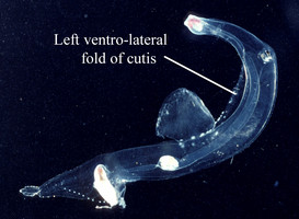
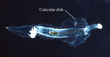
Figure. Left: View of left side of body in Pterotrachea hippocampus. Right: dorsal view of P. scutata. ©
- In dorsal view, eye shape either rectangular (P. coronata and P. scutata) or narrowly to broadly triangular, as a function of age (P. hippocampus)
Click on an image to view larger version & data in a new window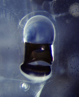
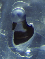
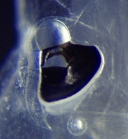
Figure. Eyes of Pterotrachea scutata (left) and P. hippocampus (center and right). ©
- Sucker medial on ventral surface of swimming fin; present only in males
Click on an image to view larger version & data in a new window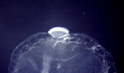
Figure. Side view of swimming fin sucker in male Pterotrachea hippocampus. ©
- Tail terminates in two flattened lobes (see title illustration), between which an elongate filament emerges (Lalli and Gilmer, 1989). The filament is present in both sexes and can extend up to 60 mm in length in Pterotrachea coronata. It is comprised of pigmented nodules (see figure below) that have been observed in the field (R. Gilmer; in Lalli and Gilmer, 1989) to expand and contract rapidly if the animal is disturbed. The tail filament is normally missing in preserved animals, undoubtedly lost during net capture.
Click on an image to view larger version & data in a new window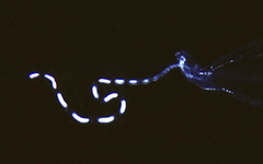
Figure. Tail filament of Pterotrachea coronata, viewed from right side. ©
- Shell present only in larvae (see below) and cast off after metamorphosis
- Tentacles absent in both sexes
- Radula with central rachidian teeth that are polycuspid, with a large, pointed median cusp and multiple side cusps. Lateral and marginal teeth are monocuspid
Click on an image to view larger version & data in a new window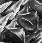
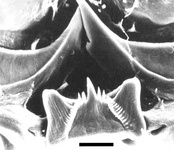
Figure. Scanning electron micrographs of central rachidian teeth in Pterotrachea hippocampus. Photograph on left modified from Thiriot-Quiévreux (1973, Fig. 1I. © 1973 C. Thiriot. Photograph on right modified from Ricther and Seapy (1999, Fig. 4D). © G. Richter.
- Larvae
- Three larvae, designated larvae 1, 2 and 3 by Richter (1968), are recognized in the genus Pterotrachea. The shells of the three larvae all consist of about 2 and 1/2 whorls, but are distinguished by the following: in larva 1, the surface is smooth and the last whorl separates from the inner whorls; in larva 2, the last whorl does not separate from the preceding whorls, the shell is thin and transparent, and the surface is tranversed by about 30 ridges; and, in larva 3 the shell is coiled and has a shape similar to larva 2, but the surface is smooth.
Click on an image to view larger version & data in a new window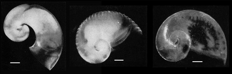
Figure. The three larval types in the genus Pterotrachea: larva 1 (left), larva 2 (center), and larva 3 (right). Plate created from photographs in Richter (1968, Figs. 19, 20, and 21). All scale bars = 100 µm. © 1968 G. Richter
- In her review of the Heteropoda, Thiriot-Quiévreux (1973) confirmed the identifications by Richter of the three larval types in the Mediterranean Sea, and she noted that larva 2 is the most frequently encountered of the three types.
- The third of the three larval types is distinguished immediately by its velum. In larvae 1 and 2 the velum consists of four long and slender lobes of equal length (see first figure below), which are similar to the velar lobes in the larva of Firoloida desmarestia. While three of the velar lobes in larva 3 are like those in the other two larvae, the left frontal lobe is widened and winglike, and has a brown stain in the center of the tip (Richter, 1968; see second figure below). Click on an image to view larger version & data in a new window
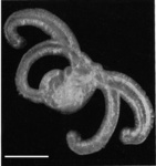
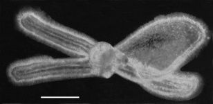
Figure. Photographs of type 2 (left) and type 3 (right) larvae of Pterotrachea. Modified from Richter (1968: Figs. 22 and 23). Scale bars = 50 µm © 1968 G. Richter
- Because the larval shell is shed following metamorphosis and early post-metamorphic development of the three species appears to be very similar, tentative identifications of the larvae at the species level are speculative (see Comments section below).
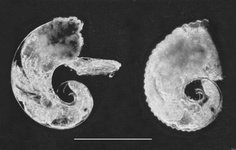
Figure. Photographs of Pterotrachea larva 1 (left) and larva 2 (right); note bodies retracted into the shells. Scale bar = 500 µm. Modified from Thiriot-Quiévreux (1973, Fig. 7A,B). © 1973 Catherine Thiriot
Comments
Krohn (1860) was the first to describe and illustrate the two larval types of Pterotrachea that Richter (1968) later termed Pterotrachea larvae 1 and 2.
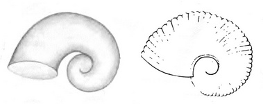
Figure. Two larval types of Pterotrachea, viewed from the left side. Modified from Krohn (1860, Figs. 22, 24). © 1860 A. Krohn
From western Mediterranean waters, Franc (1948) described a larva whose shell corresponded with that of Krohn's (1860) second larval type, and which he identified as Firola (Pterotrachea) coronata; although no justification was given for the species identification.
The two larval types of Krohn (1860) are also known from fossils, dated to the Quaternary Period (scanning electron micrographs below courtesy of Arie Janssen).
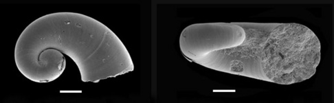
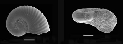
Figure. Scanning electron micrographs of fossil Pterotrachea larva 1 (top pair of photographs) and larva 2 (Bottom photographs), viewed from the right side (left) and aperture (right). Specimens from benthic core samples (700 m depth) in the Eastern Mediterranean and dated to the Quaternary Period. Scale bars = 100 µm. © 2007 Arie W. Janssen
Richter (1968) made tentative species identifications of the three larval types based on observations of early development following metamorphosis. Only the early stages of eye formation provided a clue to species identity; the shape of the visceral nucleus, important in distinguishing P. coronata, was not discernable at this early stage of development. After metamorphosis of larva 1, the eye had a broad base, which Richter indicated would correspond with that seen in adult P. hippocampus. Following metamorphosis of larva 2, the base of the eye was found to be similar to the preceding species, but narrower; which Richter indicated was like that in adult P. minuta. Lastly, after metamorphosis in larva 3 the eye base was narrow, as seen in adult P. coronata or P. scutata. Given the determination that specimens previously identified as P. minuta are actually young P. hippocampus (Seapy, 1985, 2000), the tentative identity of larva 2 as P. minuta must be revisited. Richter's hypothesized identification of larva 1 as P. hippocampus is most probably correct, which leaves the other two larval types as either P. coronata or P. scutata. Unfortunately, the eyes of the latter two species are very similar in shape and appearance (see Seapy, 1985; Figs. 3F,G).
The three species in the genus Pterotrachea can be distinguished by the following:
| Species | Maximal body length | Cutis thckened on trunk anterior to swimming fin | Eye shape in dorsal view | Eye length to retinal width ratio (mean and range)* | Visceral nucleus shape | Visceral nucleus length to width ratio (mean and range)* |
|---|---|---|---|---|---|---|
| P. hippocampus | 80 mm | as ventro-lateral folds, forming a ventral bib | narrowly to broadly triangular | 1.3 (1.1-1.6) | short; tear-drop | 3.0 (2.0-3.7) |
| P. coronata | 330 mm | as ventro-lateral folds, forming a ventral bib | rectangular | 2.2 (1.9-2.5) | tall; narrow tear-drop | 5.6 (4.1-7.2) |
| P. scutata | 200 mm | as a laterally projecting, flattened oval disk | rectangular | 2.2 (2.1-2.4) | short; tear-drop | 3.0 (2.0-3.9) |
*Ratios (means and ranges) from Seapy (1985, Figs. 4 and 5). Based on regression analysis, the eye length to retinal width ratios of P. coronata and P. hippocampus decreased with increasing body length. A similar pattern was seen, but was less pronounced, for the ratio of visceral nucleus length to width in P. hippocampus. Remaining ratios showed essentially no change with increasing body length.
References
Franc, A. 1948. Veligeres et mollusques gasteropodes des Baies D'Alger et de Banyuls. Journal de Conchyliologie (Paris, France). (4)88(41): 13-42.
Krohn, 1860. Beitrage zur Entwicklungsgeschichte der Pteropoden und Heteropoden. 43 S., 2 Tafeln, Leipzig.
Lalli, C. M. and R. W. Gilmer. 1989. Pelagic snails. The biology of holoplanktonic gastropod mollusks. Stanford Unive. Press, Stanford, pp. 1-259.
Richter, G. 1968. Heteropoden und Heteropodenlarven im Oberflachenplankton des Golfs von Neapel. Pubblicazioni della Stazione Zoologica di Napoli 36: 346-400.
Richter, G. and R. R. Seapy. 1999. Heteropoda, pp. 621-647. In: D. Boltovskoy (ed.), South Atlantic Zooplankton. Leiden: Backhuys Publ.
Seapy, R.R. 1985. The pelagic genus Pterotrachea (Gastropoda: Heteropoda) from Hawaiian waters: a taxonomic review. Malacologia 26(1-2): 125-135.
Seapy, R.r. 2000. Species discrimination among pelagic heteropods: resolution of the Pterotrachea hippocampus - P. minuta problem. Journal of Molluscan Studies 66: 99-117.
Thiriot-Quievreux, C. 1973. Heteropoda. Oceanography and Marine Biology, an Annual Review 11: 237-261.
Title Illustrations
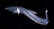
| Scientific Name | Pterotrachea coronata and Pterotrachea |
|---|---|
| Location | Hawaiian Islands |
| Specimen Condition | Live Specimen |
| Sex | Female |
| Life Cycle Stage | adult |
| View | left side |
| ToL Image Use |
 This media file is licensed under the Creative Commons Attribution-NonCommercial License - Version 3.0. This media file is licensed under the Creative Commons Attribution-NonCommercial License - Version 3.0.
|
| Copyright |
©

|
About This Page
California State University, Fullerton, California, USA
Correspondence regarding this page should be directed to Roger R. Seapy at
Page copyright © 2007
 Page: Tree of Life
Pterotrachea .
Authored by
Roger R. Seapy.
The TEXT of this page is licensed under the
Creative Commons Attribution License - Version 3.0. Note that images and other media
featured on this page are each governed by their own license, and they may or may not be available
for reuse. Click on an image or a media link to access the media data window, which provides the
relevant licensing information. For the general terms and conditions of ToL material reuse and
redistribution, please see the Tree of Life Copyright
Policies.
Page: Tree of Life
Pterotrachea .
Authored by
Roger R. Seapy.
The TEXT of this page is licensed under the
Creative Commons Attribution License - Version 3.0. Note that images and other media
featured on this page are each governed by their own license, and they may or may not be available
for reuse. Click on an image or a media link to access the media data window, which provides the
relevant licensing information. For the general terms and conditions of ToL material reuse and
redistribution, please see the Tree of Life Copyright
Policies.
- First online 02 November 2007
- Content changed 13 August 2008
Citing this page:
Seapy, Roger R. . 2008. Pterotrachea . Version 13 August 2008 (under construction). http://tolweb.org/Pterotrachea/28737/2008.08.13 in The Tree of Life Web Project, http://tolweb.org/


