


Pterotrachea coronata
Roger R. Seapy

Introduction
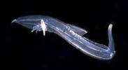
Pterotrachea coronata attains the largest size (to 330 mm) among the Pterotracheidae and has the most streamlined body shape. The proboscis is long and pointed, terminating in a small buccal mass. The trunk is long and slender, with ventro-lateral folds of cutis anterior to the swimming fin that form a concave bib. The visceral mass is long and slender (the highest length to width ratio in the genus), and the basal part of its length is deeply imbedded in the posterior end of the trunk. The tail is large and laterally flattened, creating a broad surface area that aids in burst swimming, attributable to strong side-to-side flexion of the trunk and tail. During burst swimming, the proboscis is tucked into a ventral groove in the anterior one-half of the trunk created by the ventro-lateral folds of cutis. When viewed dorsally the eyes have a rectangular shape with a retinal base somewhat wider than the lens (very similar to the eye in P. scutata). A fin sucker is located on the mid-ventral surface of the swimming fin.
Brief Diagnosis:
A species in the genus Pterotrachea with the following characteristics:
- The most streamlined body among the pterotracheids
- Proboscis slender and elongate, terminating in a small buccal mass
- During burst swimming, proboscis is tucked into the ventral groove formed by the ventro-lateral folds of cutis
- Burst swimming is the result of side-to-side body flexion, involving the long, narrow trunk and the large, laterally flattened tail
- Eyes rectangular in dorsal view, with the retinal base somewhat wider than the lens
- Swimming fin sucker only in males
Characteristics
- Body morphology
- Body length greatest among the pterotracheids (to 330 mm)
- Streamlined body shape; trunk elongate and cylindrical, tail large and laterally flattened (see title illustration)
- Proboscis long, slender and cylindrical, tapering to a small buccal mass (see title illustration)
- Eye shape rectangular in dorsal view, with retinal base somewhat wider than the lens; very similar in appearance to that in P. scutata. In Hawaiian P. coronata the average eye length to width ratio = 2.2, although the ratio decreases markedly from 2.5 in small to 1.9 in large individuals (Seapy, 1985)Click on an image to view larger version & data in a new windowClick on an image to view larger version & data in a new window

Figure. Left eye of Pterotrachea coronata. ©
- Visceral nucleus long and narrow; the highest among the Pterotracheidae (see the figure below and the title illustration). In Hawaiian P. coronata the ratio of length to width averages 5.6 and ranges from 4.1-7.2 (Seapy, 1985).
- Visceral nucleus imbedded in the cutis, with only the tapered, pointed end extending above the posterior end of the trunkClick on an image to view larger version & data in a new windowClick on an image to view larger version & data in a new window
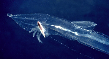
Figure. Posterior portion of body in a female Pterotrachea coronata, viewed from the left side. Note the long egg string that extends below the visceral nucleus toward the lower right. Note also (by clicking on the image for an enlarged view) the linear arrangement of the devoloping embryos in the transparent mucus sleeve. ©
- Swimming fin closer to the visceral nucleus than to the head, with the anterior end of fin just anterior to the midpoint of the trunk (see title illustration)
- Sucker located medially on ventral margin of the swimming fin; present only in males
Click on an image to view larger version & data in a new window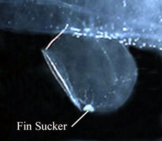
Figure. Swimming fin and sucker in a male Pterotrachea coronata, viewed from right side.©
- Tail terminates in a pair of small, laterally-flattened lobes, between which emerges an elongate filament that is not always present in the field and is invariably missing in net-captured individualsClick on an image to view larger version & data in a new window
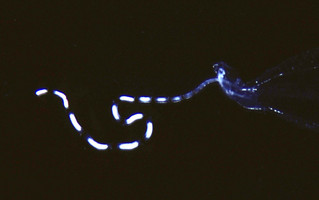
Figure. Tail filament of Pterotrachea coronata, viewed from right side. ©
- Shell present only in larvae and is lost following metamorphosis
- Radular morphology not described, but presumably similar to that in the other species in the genus (see Pterotrachea page)
- Larva unknown, but probably is represented by either larva 2 or larva 3 of Richter (1968); see Pterotrachea page.
References
Lalli, C. M. and R. W. Gilmer. 1989. Pelagic snails. The biology of holoplanktonic gastropod mollusks. Stanford Unive. Press, Stanford, pp. 1-259.
Richter, G. and R. R. Seapy. 1999. Heteropoda, pp. 621-647. In: D. Boltovskoy (ed.), South Atlantic Zooplankton. Leiden: Backhuys Publ.
Seapy, R.R. 1985. The pelagic genus Pterotrachea (Gastropoda: Heteropoda) from Hawaiian waters: a taxonomic review. Malacologia 26(1-2): 125-135.
Title Illustrations
| Scientific Name | Pterotrachea coronata and Pterotrachea |
|---|---|
| Location | Hawaiian Islands |
| Specimen Condition | Live Specimen |
| Sex | Female |
| Life Cycle Stage | adult |
| View | left side |
| ToL Image Use |
 This media file is licensed under the Creative Commons Attribution-NonCommercial License - Version 3.0. This media file is licensed under the Creative Commons Attribution-NonCommercial License - Version 3.0.
|
| Copyright |
©

|
About This Page
California State University, Fullerton, California, USA
Correspondence regarding this page should be directed to Roger R. Seapy at
Page copyright © 2007
 Page: Tree of Life
Pterotrachea coronata .
Authored by
Roger R. Seapy.
The TEXT of this page is licensed under the
Creative Commons Attribution License - Version 3.0. Note that images and other media
featured on this page are each governed by their own license, and they may or may not be available
for reuse. Click on an image or a media link to access the media data window, which provides the
relevant licensing information. For the general terms and conditions of ToL material reuse and
redistribution, please see the Tree of Life Copyright
Policies.
Page: Tree of Life
Pterotrachea coronata .
Authored by
Roger R. Seapy.
The TEXT of this page is licensed under the
Creative Commons Attribution License - Version 3.0. Note that images and other media
featured on this page are each governed by their own license, and they may or may not be available
for reuse. Click on an image or a media link to access the media data window, which provides the
relevant licensing information. For the general terms and conditions of ToL material reuse and
redistribution, please see the Tree of Life Copyright
Policies.
- First online 18 August 2008
- Content changed 18 August 2008
Citing this page:
Seapy, Roger R. . 2008. Pterotrachea coronata . Version 18 August 2008 (under construction). http://tolweb.org/Pterotrachea_coronata/28738/2008.08.18 in The Tree of Life Web Project, http://tolweb.org/


