


Pterotrachea coronata
Roger R. Seapy
Introduction
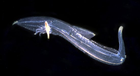
Pterotrachea coronata attains the largest size (to 330 mm) among the Pterotracheidae and has the most streamlined body shape. The proboscis is long and pointed, terminating in a small buccal mass. The trunk is long and slender, with ventro-lateral folds of cutis anterior to the swimming fin that form a concave bib. The visceral mass is long and slender (the highest length to width ratio in the genus), and the basal part of its length is deeply imbedded in the posterior end of the trunk. The tail is large and laterally flattened, creating a broad surface area that aids in burst swimming, attributable to strong side-to-side flexion of the trunk and tail. During burst swimming, the proboscis is tucked into a ventral groove in the anterior one-half of the trunk created by the ventro-lateral folds of cutis. When viewed dorsally the eyes have a rectangular shape with a retinal base somewhat wider than the lens; shape similar to that in P. scutata. A small fin sucker is located on the mid-ventral surface of the swimming fin in males only.
Brief Diagnosis:
A species in the genus Pterotrachea with the following characteristics:
- The most streamlined body among the pterotracheids
- Proboscis slender and elongate, terminating in a small buccal mass
- During burst swimming, proboscis is tucked into the ventral groove formed by the ventro-lateral folds of cutis
- Eyes rectangular in dorsal view, with the retinal base somewhat wider than the lens
- Swimming fin sucker only in males
- Burst swimming is the result of side-to-side body flexion, involving the long, narrow trunk and the large, laterally flattened tail
Characteristics
- Body morphology
- Body length greatest among the pterotracheids (to 330 mm)
- Streamlined body shape; trunk elongate and cylindrical, tail large and laterally flattened (see title illustration)
- Proboscis long, slender and cylindrical, tapering to a small buccal mass (see title illustration)
- Eye shape rectangular in dorsal view, with retinal base somewhat wider than the lens; very similar in appearance to that in P. scutata. In Hawaiian P. coronata the average eye length to width ratio = 2.2, decreasing from 2.5 in small to 1.9 in large individuals (Seapy, 1985)Click on an image to view larger version & data in a new window
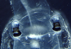
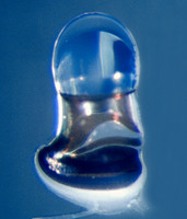
Figure. Dorsal views of the head region (left) and a dissected right eye (right) in Pterotrachea coronata. ©
- Visceral nucleus long and narrow (see the figure below and the title illustration). The shape contrasts markedly with the shorter, tear-drop shape of the visceral nucleus in P. scutata and P. hippocampus (see their respective pages). In Hawaiian P. coronata the ratio of length to width averages 5.6 and ranges from 4.1-7.2 (Seapy, 1985).
- Visceral nucleus imbedded in the cutis, with only the tapered, pointed end extending above the posterior end of the trunkClick on an image to view larger version & data in a new window

Figure. Posterior portion of body in a female Pterotrachea coronata, viewed from the left side. Note the gills projecting dorsally and the long egg string that extends below the visceral nucleus toward the lower right. Note also (by clicking on the image for an enlarged view) the linear arrangement of the devoloping embryos in the transparent mucus sleeve. ©
- Swimming fin closer to the visceral nucleus than to the head, with the anterior end of fin just anterior to the midpoint of the trunk (see title illustration)
- Small sucker located medially on ventral margin of the swimming fin and present only in males Click on an image to view larger version & data in a new window
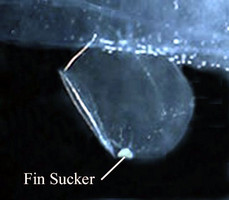
Figure. Swimming fin and sucker in a male Pterotrachea coronata, viewed from right side.©
- Tail terminates in a pair of small, laterally-flattened lobes (see title illustration), between which emerges an elongate filament that is not always present in the field and is usually missing in net-captured individuals Click on an image to view larger version & data in a new window
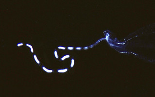
Figure. Tail filament of Pterotrachea coronata, viewed from right side. ©
- Shell present only in larvae and is lost following metamorphosis
- Larva unknown, but probably is represented by either larva 2 or larva 3 of Richter (1968); see Pterotrachea page.
- Radular morphology similar to that in the other species in the genus, with a median cusp that is conspicuously broader and longer than the lateral cusps (see Pterotrachea page).
Comments:
Swimming behavior has been observed in the field (Gilmer, in Lalli and Gilmer, 1989). Animals were reported to exhibit different behavior during daytime and nighttime hours. During the day, animals swam with their ventral side directed upward (see title illustration), which would facilitate visual location of prey silhouetted against the lighted surface waters. Active swimming occurred when animals were observed pursuing prey or when they were disturbed by scuba divers (and, presumably, "natural" predators). At other times they appeared to be neutrally buoyant, floating curled into loose balls. At night, animals also appeared to be neutrally buoyant, but were oriented with their ventral sides directed downward with or without slow undulation of the swimming fin. In this position, animals hypothetically could locate bioluminescent prey beneath them.
Pterotrachea coronata was studied by Denton and Shaw (1961) to determine how these animals are able to achieve neutral buoyancy. The authors found that body fluids obtained by centrifugation of the gelatinous tissues had a lower density than sea water, attributable to the replacement of heavier sulfate by lighter chloride ions. The sulfate ion concentration in the body fluids was shown to be about three-fourths that in sea water.
References
Denton, E. J. and T. I. P. Shaw. 1961. The buoyancy of gelatinous marine animals. Journal of Physiology, London 161: 14P-15P (Proceedings).
Lalli, C. M. and R. W. Gilmer. 1989. Pelagic snails. The biology of holoplanktonic gastropod mollusks. Stanford Unive. Press, Stanford, pp. 1-259.
Richter, G. 1968. Heteropoden und Heteropodenlarven im Oberflachenplankton des Golfs von Neapel. Pubblicazioni della Stazione Zoologica di Napoli 36: 346-400.
Richter, G. and R. R. Seapy. 1999. Heteropoda, pp. 621-647. In: D. Boltovskoy (ed.), South Atlantic Zooplankton. Leiden: Backhuys Publ.
Seapy, R.R. 1985. The pelagic genus Pterotrachea (Gastropoda: Heteropoda) from Hawaiian waters: a taxonomic review. Malacologia 26(1-2): 125-135.
Title Illustrations

| Scientific Name | Pterotrachea coronata and Pterotrachea |
|---|---|
| Location | Hawaiian Islands |
| Specimen Condition | Live Specimen |
| Sex | Female |
| Life Cycle Stage | adult |
| View | left side |
| ToL Image Use |
 This media file is licensed under the Creative Commons Attribution-NonCommercial License - Version 3.0. This media file is licensed under the Creative Commons Attribution-NonCommercial License - Version 3.0.
|
| Copyright |
©

|
About This Page
California State University, Fullerton, California, USA
Correspondence regarding this page should be directed to Roger R. Seapy at
Page copyright © 2007
 Page: Tree of Life
Pterotrachea coronata .
Authored by
Roger R. Seapy.
The TEXT of this page is licensed under the
Creative Commons Attribution License - Version 3.0. Note that images and other media
featured on this page are each governed by their own license, and they may or may not be available
for reuse. Click on an image or a media link to access the media data window, which provides the
relevant licensing information. For the general terms and conditions of ToL material reuse and
redistribution, please see the Tree of Life Copyright
Policies.
Page: Tree of Life
Pterotrachea coronata .
Authored by
Roger R. Seapy.
The TEXT of this page is licensed under the
Creative Commons Attribution License - Version 3.0. Note that images and other media
featured on this page are each governed by their own license, and they may or may not be available
for reuse. Click on an image or a media link to access the media data window, which provides the
relevant licensing information. For the general terms and conditions of ToL material reuse and
redistribution, please see the Tree of Life Copyright
Policies.
- First online 18 August 2008
- Content changed 17 September 2008
Citing this page:
Seapy, Roger R. . 2008. Pterotrachea coronata . Version 17 September 2008 (under construction). http://tolweb.org/Pterotrachea_coronata/28738/2008.09.17 in The Tree of Life Web Project, http://tolweb.org/


