A ventral view of this brain and the optic lobes can be found here.
J. Z. Young, the leading authority on the cephalopod nervous system in the latter half of the 20th century, divided the cephalopod brain into two regions: the supraesophageal mass and the subesophageal mass. These regions are joined laterally by two regions, the basal lobes and the dorsal magnocellular lobes. This arrangement suggest the ancestral brain was formed from two partially-circumesophageal cords that included the middle and posterior subesophageal masses and that these were fused dorsally with a third cord, the supraesophageal mass. The supra- and subesophageal masses further connected by the cerebro-brachial connective that runs between the anterior regions. The latter connection involves the anterior subesophageal mass and the superior buccal lobes. In decapodiforms these lobes are well removed from the rest of the brain and suggests that either they were not part of the original circumesophageal chords or formed a separate more anterior cord.

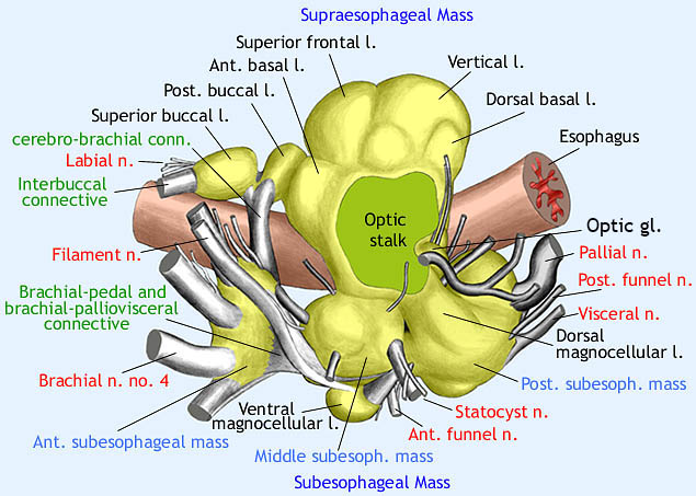
Figure. Lateral view of the brain of Vampyroteuthis infernalis reconstructed from histological sections. Drawing modified from Young (1967).
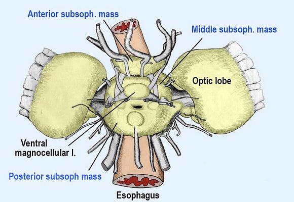
Figure. Ventral view of the brain of Vampyroteuthis infernalis reconstructed from histological sections. Drawing by R. Young.
The masses of the more posterior portion of the brain are joined laterally by the complex basal lobes and the dorsal magnocellular lobes. The separate nature of the lobes that join the supra- and subesophageal masses and, therefore, evidence for two rather than one ancestral cords, is apparent from the presence of a fissure between these regions (see dark area in drawing of Vampyroteuthis brain) which completely separates them at one point and a ventral fissure between the middle and posterior subesophageal masses which forms a path for the cephalic arteries (not visible in this drawing).
In Nautilus, the three cords that comprise the brain are more apparent. The two ventral cords partially fuse prior to their joining the dorsal cord. This fusion is apparent by their slight more dorsal separation where a fissure (J. Z. Young, 1965) separates the magnocellular lobe, at one point, from a more anterior brachial lobe (comparable to the anterior and middle subesophageal masses of coleoids) and therefore lies at the same point as the fissure in Vampyroteuthis. In Nautilus the two ventral cords, while joined laterally, are entirely separate over most of their length ventrally.


