On this page we include three families (all oegopsids), the Onychoteuthidae (except Ancistroteuthis), Enoploteuthidae and the Lycoteuthidae. Several taxa included in the Atypical Beaks page have beaks with strong similarities to Tripartite beaks.
ORDER OEGOPSIDA
ONYCHOTEUTHIDAE: 7 genera
UPPER BEAK CHARACTERISTICS
- Step: Present.
- Shoulder Blade: Thick, located in Middle Part of Shoulder; mostly medial to an Outer Cover with slightly translucent whitish/yellowish pigment which is thickest at its dorsal end. The Tripartite Shoulder Blade is not the same structure as the Bipartite Shoulder Blade.
- Shoulder Blade: Complex; Consists of two Components (Inner and Outer). Components form simultaneously as single unit with bend in midline (bend defines Components), and independently of the pigmented Lateral Wall. The Outer Component is continuous posteriorly with the Free Shoulder and the Inner Component extends posteriorly onto the Sessile Shoulder.
- Shoulder Blade: Anterior edge with S-shape in profile view.
- Origin of Shoulder Blade: Unknown. Possibly derived from Bridge.
- Hyaline Matrix: Forms a layer medial to the Shoulder Blade.
- Palatoshoulder Ridge: Present. Reduced (i.e., less extensive than in Bipartite beaks); without a free edge.
- Pigmentation of posterior surface of Hyaline Matrix: Posterior surface of Shoulder Blade forms most of the posterior pigmentation of the Shoulder. It extends, during ontogeny, over posterior surface of Hyaline Matrix. This pigmentation has a sagittate "leaf-shape" appearance with a grove near the midline which corresponds to the junction of the Free and Sessile Shoulders.
- Yellow line: Absent. Dorsal side of Outer Part of Shoulder can look somewhat similar to Yellow Line.
Phylogenetic relationships. Morphology has not provided convincing evidence of familial relationships for the Onychoteuthidae. In Lindgren's (2010) molecular study, the Likelihood analysis found the Onychoteuthidae sister to the Gonatidae (with good support); the Bayesian analysis found the Onychoteuthidae sister to the Gonatidae (with very good support) and these two families in turn were sister to the Ommastrephidae + Cranchiidae (with very good support); the parsimony analysis found the familial relationships of the Onychoteuthidae unresolved. The Lindgren et al. (2012) study found the Onychoteuthidae sister to the Pyroteuthidae + Enoploteuthidae (with weak support); these three families were sister to the Gonatidae + Brachioteuthidae (with weak support) and these five families were sister to Cycloteuthis (with weak support).

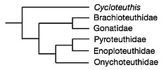
Figure. Relationships of the Onychoteuthidae. Modified from Lindgren et al., 2012.
Onychoteuthidae: Structure
- EXTERNAL FEATURES
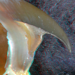
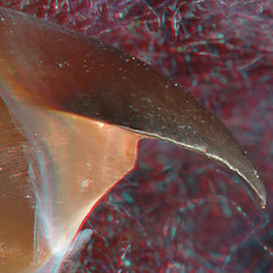
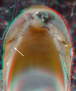
- The major parts. The Shoulder Blade (white arrow in image below) is a thick, prominent structure that occupies middle of the shoulder rather than its lateral surface as seen in Bipartite beaks. A translucent Outer Cover is present lateral to the Shoulder Blade. A Hyaline Matrix is present on the medial side of the Shoulder Blade and medial to the Hyaline Matrix is a reduced Palatoshoulder Ridge. The Shoulder Blade is, apparently, not homologous to that of the Bipartite beak and may be a derivative of the Bridge. Nevertheless, we use the same formal name, Shoulder Blade, for this heavily pigmented structure of the shoulder. The evolutionary relationship of the Outer Layer to the Outer Part of the Bipartite beak is unknown.
Click on an image to view larger version & data in a new window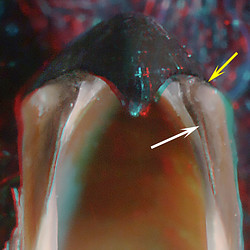
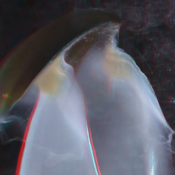
Figure Front view of the upper beak of Onychoteuthis compacta, 90 mm ML, 2.7 mm URL. White arrow points to Shoulder Blade; yellow arrow points to the "step." Note the distinct curve in the Shoulder Blade (see cross-sectional cut below).
- The Step. A Step is present between the Shoulder Blade and the jaw edge (see yellow arrows in images above and below). That is, the Outer Layer is partially or entirely absent from the Shoulder Blade here and the the Jaw Edge Extension begins well posterior to the junction of the Shoulder Blade and Palate.
Click on an image to view larger version & data in a new window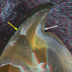
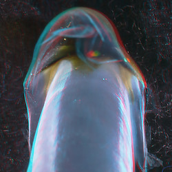
Figure. Oblique view, same beak as above. The left arrow indicates the Step and the right arrow indicates the forward curving extension of the Shoulder Blade onto the Palate.
- The Shoulder Blade. In profile (= side) view the Shoulder Blade forms most of the anterior (= leading) edge of the Shoulder. The Blade is mostly convex (sometimes as a distinct tooth) but as it approaches the Jaw Edge, the leading edge bends forward (= anteriorly) onto the Palate. This S-shape sometimes is not obvious in young beaks but becomes accentuated with growth.
Click on an image to view larger version & data in a new window
Figure. Side view, same beak as above. Arrow points to the anterior curvature of the Shoulder Blade onto the Palate.
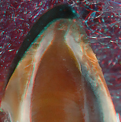
- The Outer Cover of the Shoulder. This lies lateral to the Shoulder Blade. In beaks without heavy pigmentation the Layer has a slightly translucent whitish or yellowish pigmentation. It is thickest, and therefore more easily seen, at the top (= dorsal side) of the shoulder adjacent to the jaw-edge extension (arrow). This should not be confused with the very different Yellow Line of ommastrephids and their close relatives. Often most of this yellowish material seems to have a very thin cover which may, or may not, be the homologue of the true shoulder blade seen in Bipartite beaks. The Outer Cover is generally worn away at the leading edge of the Shoulder Blade and in older beaks it loses its distinctive appearance as it becomes heavily pigmented. In this latter state, the Outer Cover can look much like the Shoulder Blade of Bipartite beaks. Click on an image to view larger version & data in a new window
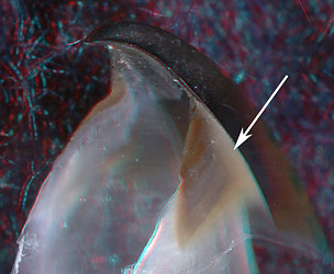
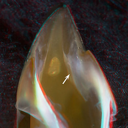
Figure. Oblique view of the anterior part of the upper beak of Onychoteuthis borealijaponica, 80 mm ML, 2.6 mm URL. Arrow indicates whitish or yellowish hyaline material of the Outer Layer of the Shoulder.
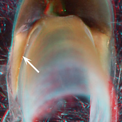
- The Palatoshoulder Ridge. This Ridge differs from that of the Bipartite Shoulder in that it is not the entire Lateral Wall in this region, but a relatively thin and narrow, inner, pigmented layer of the Lateral Wall that has separated from the main Lateral Wall. It is not easily recognizable until the Lateral Wall approaches full pigmentation. The Ridge is well posterior to the leading edge of the Shoulder Blade and is further separated from it medially by the Hyaline Matrix. In heavily pigmented beaks the Hyaline Matrix shrinks greatly and the Palatoshoulder Ridge abuts the Shoulder Blade, sometimes with a superficial residue of white Hyaline Matrix between the two, but the Ridge remains posterior to the leading edge of the Shoulder Blade.
Click on an image to view larger version & data in a new window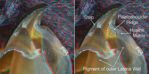
Figure. O. compacta, 90 mm ML, 2.7 mm URL, oblique views. Left - Beak without any interfering drawing or text. Right - Palatoshoulder Ridge outlined with a solid line along its anterior and ventral edges and a dashed line indicating where the Ridge fuses (separates from) the rest of the Lateral Wall.
-
Pigmentation of the posterior surface of the Shoulder. The Shoulder Blade pigmentation appears early in ontogeny on the posterior surface of the Shoulder with a "leaf" shape (i.e., a sagittate leaf) with a groove near the center. One side of the "leaf" lies on the inner surface of the Free Shoulder and the other on the Sessile Shoulder. The groove between them marks the bend that defines the two parts of the Shoulder Blade (see Strructure below). The Shoulder Blade appears to provide most of the pigmentation of the posterior surface of the Shoulder as the squid grows.
Click on an image to view larger version & data in a new window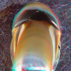
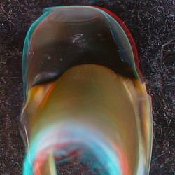
Figure. Posterior view, Onychoteuthis borealijaponica, immature female, 110 mm ML, 2.8 mm URL. Arrow points to leaf-shaped pigment of the posterior surface of the Shoulder Blade.
- The major parts. The Shoulder Blade (white arrow in image below) is a thick, prominent structure that occupies middle of the shoulder rather than its lateral surface as seen in Bipartite beaks. A translucent Outer Cover is present lateral to the Shoulder Blade. A Hyaline Matrix is present on the medial side of the Shoulder Blade and medial to the Hyaline Matrix is a reduced Palatoshoulder Ridge. The Shoulder Blade is, apparently, not homologous to that of the Bipartite beak and may be a derivative of the Bridge. Nevertheless, we use the same formal name, Shoulder Blade, for this heavily pigmented structure of the shoulder. The evolutionary relationship of the Outer Layer to the Outer Part of the Bipartite beak is unknown.
- INTERNAL STRUCTURE: Longitudinal cuts through the Shoulder.
- A longitudinal cut from the beak of a young Onychoteuthis borealijaponica, 110 mm ML, 2.8 mm URL. Click on an image to view larger version & data in a new window

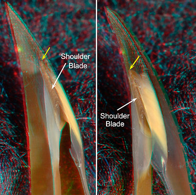
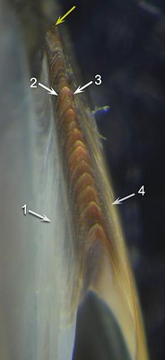
Figure. Onychoteuthis borealijaponica, immature, 110 mm ML. T0P: Side view showing where the beak was cut. BOTTOM: Left - Oral view of the cut surface of the shoulder. This image shows the medial edge of the Hyaline Matrix clearly. Middle - Oral-lateral view. Right - Photomicrograph (2D) of the cut Shoulder, oral view taken perpendicular to the cut surface.
ARROWS: 1 - Hyaline Matrix. 2 - Inner Component of the Shoulder Blade. 3 - Outer Component of the Shoulder Blade. 4 - Outer Layer. Yellow arrows - Point to the same spot on all beaks.COMMENTS. The cut shows the characteristic structure of the Shoulder Blade with a bend that separates its Inner and Outer Components. The bend is slightly lateral to the midline making the Outer Component a bit narrower than the Inner Component. The Outer Layer is easily recognized by its yellow color. A thick Hyaline Matrix covers the posterior 2/3 of the Blade on its medial side. The pigment of the posterior Lateral Wall ends where the thick Hyaline Matrix begins. The Palatoshoulder Ridge has not yet developed (ie., the pigmented Lateral Wall terminates at the Shoulder both posteriorly and dorsally) in this young beak.
- Two cuts from a much older, but still young Onychoteuthis borealijaponica, 175 mm ML, ~4.4 mm URL.
In preparation, the beak was first cut approximately in the midline to make cuts through the Shoulder easier. Next two cuts were made on one side to examine the Shoulder structure. Cut 1 was a longitudinal cut that was parallel to the Jaw-edge Extension. Cut 2 was more dorsal and oblique and passed through the reduced Palatoshoulder Ridge. The beak often shatters at bit during cutting rather than providing a simple, smooth cut. The top two tiers show the cuts in 3D images and the bottom tier shows the same cuts in 2D from photomicrographs with higher resolution.
Click on an image to view larger version & data in a new window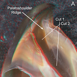
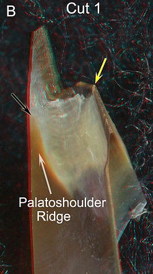
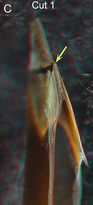
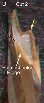

Figure. Onychoteuthis borealijaponica, immature female, 175 mm ML, ~4.4 mm URL. 3D images to aid in viewer orientation. A - Oblique view of the uncut beak showing the direction of the two cuts. B, C - Cut 1 images show two slightly different views (B- oral-medial, C- oral) of this more ventral cut. D, E - Cut 2 images with approximately the same views as in B and C, although the cut is more dorsal and oblique. Yellow arrows - Point to the same spot on all beaks (i.e., anterior end of the cut Shoulder Blade).
Fig. F - The posterior surface of the Hyaline Matrix was fully covered by pigment (5) in Cut 1. Fig. G - This "midline cut" was accidentally well off the midline but nicely shows the Palatoshoulder Ridge arising from the Lateral Wall at the Crest. The Palatoshoulder Ridge was cut both dorsally (Fig. G) and ventrally (Fig. D). Fig. H - The Palatoshoulder Ridge clearly arises from the posterior Lateral Wall and a small portion of the posterior surface of the Hyaline Matrix remains unpigmented in this more dorsal cut.Click on an image to view larger version & data in a new window
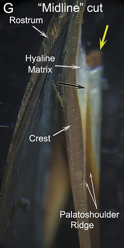

Figure. Same beak as above. Photomicrographs (2D), oral views taken perpendicular to the plane of the cuts. These 2D images show greater detail than the 3D images. F - (Cut 1) The Palatoshoulder Ridge in this cut was completely fused with the pigmented Lateral Wall and barely recognizable but this junction is not within the confines of this image. G - Image is of the beak fragment turned over. Note the position yellow arrow and black arrows; compare with similarly placed arrows in Fig. D. H - (Cut 2) Image clearly shows the Palatoshoulder Ridge branching off the Lateral Wall.
ARROWS: 1 - Hyaline Matrix. 2 - Inner component of the Shoulder Blade. 3 - Outer Component of the Shoulder Blade. 4 - Outer Layer of the Shoulder. F5 - Pigment layer across the posterior end of the Hyaline Matrix. H5 - Unpigmented gap at the posterior end of the Hyaline Matrix. Yellow arrows - Point to the same spot on all images.
COMMENTS. Except for the presence of the Palatoshoulder Ridge, the structure of the shoulder in these images (A-H) is very similar to that of the younger beak. The photomicrographs clearly show growth lines in the Shoulder Blade. Presumably with better methods, a single, continuous growth line could be followed through all the major structures of the Shoulder. We have seen the peculiar structure of the Shoulder Blade in two species of Onychoteuthis and one species of Walvisteuthis. and we presume that it is characteristic of the family, with the exception of Ancistroteuthis (see Atypical beaks).
- A longitudinal cut from the beak of a young Onychoteuthis borealijaponica, 110 mm ML, 2.8 mm URL.
- INTERNAL STRUCTURE: Cross-sectional cuts through the Shoulder.
We examined three cuts through the same beak:
Click on an image to view larger version & data in a new window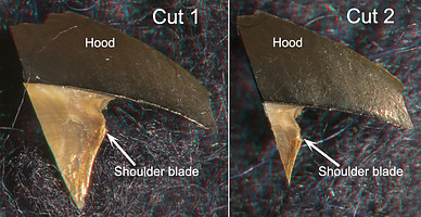
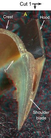
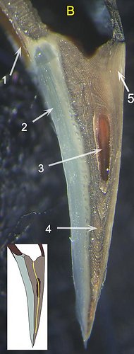

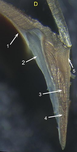
Figure. Onychoteuthis borealijaponica, immature female, 175 mm ML, ~4.4 mm URL. Two of the three parallel cross-sectional cuts through the shoulder are seen above. TOP: Side views of the beak fragments showing the position of two cuts. BOTTOM: A - Posteromedial view (3D) of cut 1 to assist in orientation. B - Photomicrograph (2D) of cut 1, Oral view taken perpendicualr to the cut, to show greater detail. B, insert - A drawing of image B with a yellow line drawn between the two components of the Shoulder Blade to show the curvature in the Blade which is often visible externally (see first image under "Basic Sructure" above). C - Posterior view (3D) of cut 2 to assist in orientation. D - Photomicrograph (2D) of cut 2, oral view, showing greater detail.
ARROWS: 1 - Palatoshoulder Ridge. 2 - Hyaline Matrix. 3 (B) - Anterior tip of Shoulder Pocket which is, also, continuous with the groove at the dorsal and posterior ends of the Shoulder Blade. 3 (D) - Just past the tip of the Shoulder Pocket. 4 - Ventral portion of the Shoulder Blade. 5 - Outer Layer of the Shoulder.
The Palatoshoulder ridge is clearly seen in B and D arising from the Crest of the Lateral Wall. This junction in Bipartite beaks approximates the point where the Lateral Wall and Bridge Join. In these cuts, however, the dorsal end of the Shoulder Blade bridges the gap between the Crest and the Hood; this is where the Bridge should be. Fig. B shows no special, dense layer in this position that could represent the Bridge. In D the gap between the Crest and Hood has narrowed and the top of the Shoulder Blade is becoming more heavily sclerotized.
Near the top of the Outer Component, the strongly curved growth lines show a specialized lateral region of the Outer Layer (B5, D5) that corresponds to the thicker yellowish area seen in a side view of the beak. The boundary between the Outer Component of the Shoulder Blade and the Outer Layer is gradual with the degree of pigmentation being the most defining feature. The Hyaline Matrix (e.g. B2) appears to be roofed dorsally mostly by the thick end of the Crest rather that by the Bridge in contrast to the condition in the Bipartite and Unipartite beaks.Cut 3 (below) was only partially successful as the lower part of the Shoulder was lost from the anterior beak fragment. As a result, we show both sides of the cut, one from the anterior fragment and one from the posterior fragment.
Click on an image to view larger version & data in a new window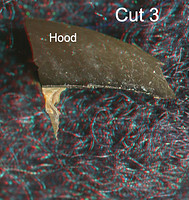


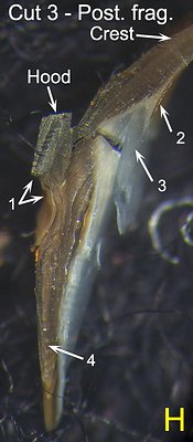
Figure. O. borealijaponica, same beak as above. TOP: Side view of the beak fragment showing the position of Cut 3. Most of the Shoulder was lost during the cut from the anterior beak fragment seen here, but was retained in the posterior fragment. BOTTOM: The two faces of the cut are shown here for clarity. Figs. E, F - Anterior fragment (note rostrum). Figs. G, H - Posterior fragment. If it were a perfect cut these sets would be mirror images of each other. E - Posterior view with Hood tilted away from viewer; 3D image for viewer orientation. F - Photomicrograph (2D) for greater detail, posterior view taken perpendicular to plane of cut. G - Anterior view, 3D image for viewer orientation. Right - Photomicrograph (2D), anterior view taken perpendicular to plane of cut.
ARROWS: 1 - Step (especially clear in E). 2 - Palatoshoulder Ridge. 3 - Hyaline Matrix. 4 - Ventral part of Shoulder Blade or its remnant.
In cut 3 the gap between the Crest and the Hood has narrowed further and the the top of the Shoulder Blade has become more sclerotized. A distinct Step is now present near the anterior end of the Shoulder. This was not apparent in the more posterior cuts. The slight convexity of the Shoulder near the Jaw-edge Extension, along with the translucency of the Outer Layer gives the impression of a longer Step when viewing the whole beak.
- CHANGES WITH GROWTH
As in all beak types, the growth stage of the beak has a large effect on how easily the beak-type is identified. Here we use taxa of the Onychoteuthidae to represent all three families.
Side
view


Onykia robusta, young male, 162 mm ML, 5.3 mm URL.
Onychoteuthis borealijaponica, immature female, 177 mm ML, 4.4 mm URL.
Onykia robsoni, male, 304 mm ML, 8.5 mm URL. The shoulder blade appears to be the first structure in the shoulder to get pigment (Left). The S-shape profile of the anterior edge of the shoulder can be recognized in all three of these images.
Oblique
view

Onykia robusta, young male, 162 mm ML, 5.3 mm URL. Onychoteuthis borealijaponica, immature female, 177 mm ML, 4.4 mm URL.
Onykia robsoni, male, 304 mm ML, 8.5 mm URL. The step in the anterior part of the shoulder also appears early. The large but young (ie, weakly sclerotized) beak fo Onykia robusta shows this clearly (Left). The Palatoshoulder Ridge is in the early states of formation in the Middle image (arrow) and often does not become a prominent part of the Shoulder until the beak is heavily pigmented (Right).
Posterior
view

Onykia robusta, young male, 162 mm ML, 5.3 mm URL. Onychoteuthis borealijaponica, immature female, 177 mm ML, 4.4 mm URL.
Onykia robsoni, male, 304 mm ML, 8.5 mm URL. The back of the shoulder in Onykia robusta (Left) and Onychoteuthis borealijaponica (Middle) show the characteristic "leaf" pattern of pigmentation. That is, a sagittate shape with a shallow groove (angle at the groove roughly between 45-90°). The "leaf" pattern, however, can be difficult to recognize in adult or near-adult squid such as Onykia robsoni (Right). Note that the Shoulder Blade arises from the middle of the Shoulder.
ENOPLOTEUTHIDAE: 4 genera
UPPER BEAK CHARACTERISTICS
- Step: Present.
- Shoulder Blade: Thick, located in Middle Part of Shoulder; Outer Cover lateral to Shoulder Blade often without translucent whitish/yellowish pigment, thickest dorsally, seen in onychoteuthids. but present in some Abraliopsis.
- Shoulder Blade: Complex; Consists of two Components (Inner and Outer). Components form simultaneously as single unit with bend in midline (bend defines Components), and independently of the pigmented Lateral Wall. Outer Component continuous posteriorly with Free Shoulder and Inner Component extends posteriorly onto Sessile Shoulder.
- Shoulder Blade: Anterior edge with S-shape in profile view.
- Origin of Shoulder Blade: Unknown. Possibly derived from Bridge.
- Hyaline Matrix: Forms layer medial to Shoulder Blade. In Enoploteuthis it covers Palatoshoulder Ridge posteriorly.
- Palatoshoulder Ridge: Present. Reduced (i.e., less extensive than in Bipartite beaks); without free edge.
- Pigmentation of posterior surface of Hyaline Matrix: Posterior surface of Shoulder Blade forms most of the posterior pigmentation of the Shoulder. It extends, during ontogeny, over posterior surface of Hyaline Matrix. This pigmentation has a sagittate "leaf-shape" appearance with a grove near the midline which corresponds to the junction of the Free and Sessile Shoulders.
- Yellow line: Absent. Dorsal side of Outer Part of Shoulder can look somewhat similar to Yellow Line in some Abraliopsis.
Phylogenetic relationships. The Enoploteuthidae is placed in the enoploteuthid families (Ancistrocheiridae, Enoploteuthidae, Pyroteuthidae and Lycoteuthidae) on the Tree of Life. There is reasonable morphological support for this grouping (Young and Harman, 1998) and especially the close relationship between the Pyroteuthidae and the Lycoteuthidae (Herring, et al., 1985). Molecular support for the group (without Ancistrocheiridae due to lack of data) has been mixed but tend to support this relationship. While Lindgren's (2010) parsimony analysis could resolve familial relationships of none the three, her Bayesian analysis placed the three families together with strong support (Enoploteuthidae + Pyroteuthidae formed a sister group to the Lycoteuthidae); her Maximum-likelihood analysis placed Pyroteuthidae and Enoploteuthidae together with good support, as did Lindgren et al. (2012) but neither of the latter analyses placed Lycoteuthidae nearby but its placement had only weak support.
Enoploteuthidae: Structure
- EXTERNAL FEATURES
We have examined many enoploteuthid beaks. With the exception of species of Enoploteuthis, the beaks are small and very difficult to examine.
Enoploteuthis higginsi, mature male, 54 mm ML, 2.5 URL. LEFT - Side view. MIDDLE - Oral-front view. Abralia astrosticta, immature female, 48 mm ML, 1.2 mm URL. RIGHT - Posterior view. LEFT - The S-shaped profile of the Shoulder Blade is apparent although the angles are rather sharp. The Outer Cover of the Shoulder Blade is not very distinct; it has no whitish/yellowish pigment concentrated dorsally. MIDDLE - The Palatoshoulder Ridge distinct. The Shoulder Blade, the Lateral Wall and the Hyaline Matrix between them are clearly seen although part of the Hyaline Matrix has become sclerotized. RIGHT - The posterior view shows the sagittate leaf-shaped pigment (arrow) of the posterior surface of the Shoulder Blade .
- INTERNAL STRUCTURE: Longitudinal cuts through the Shoulder. Click on an image to view larger version & data in a new window
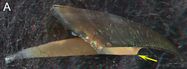
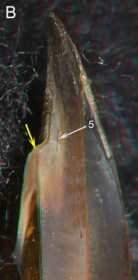

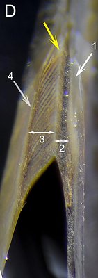
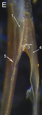
Figure. Enoploteuthis reticulata (A-D) immature female, 51 mm ML, 3.1 mm URL. A: Side view to show position of cut. B - Oral-medial view. C - Oral-lateral view.. D - Photomicrograph (2D) of cut surface of right shoulder.
Abraliopsis sp. A (E), immature female, 25 mm ML, 1.1 mm URL. Photomicrograph (2D) of cut surface of left shoulder. Arrows indicate the same structures as in adjacent photograph. The dark areas of the Hyaline Matrix are not at the cut surface but seen through the semitransparent Matrix.
ARROWS: 1 - Hyaline Matrix. 2 - Width of the Inner Component of the Shoulder Blade. 3 - Width of Outer Component of the Shoulder Blade. Note that the Outer Component has begun to sclerotize more heavily on one side to form a dark strip adjacent to the Hyaline Matrix. 4 - Outer Layer. 5 - Palatoshoulder Ridge. Yellow arrows - Points to same spot on all Enoploteuthis images. - INTERNAL STRUCTURE: Cross-sectional cuts through the Shoulder. Click on an image to view larger version & data in a new window
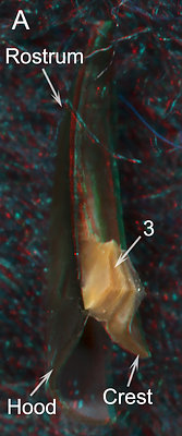
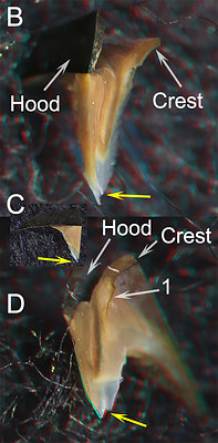
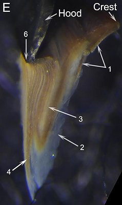
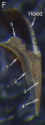
Figure. Enoploteuthis reticulata (A-E), same beak as above. A - Lateral-oral view showing both a longitudinal and a cross-sectional cut. B - Posterolateral view. C (insert) - Side view showing position of cross-sectional cut. D - Posterior-oral view. E - Photomicrograph (2D), posterior view perpendicular to plane of cut. F - Abraliopsis sp. A, same beak as above. Photomicrograph (2D), posterior view taken perpendicular to plane of cut.
ARROWS: 1 - Palatoshoulder Ridge. 2 - Hyaline Matrix. 3 - Shoulder Blade (Outer Component). 4 - Outer Layer of the Shoulder. 5 - Crest ridge. 6 - Marks the outer (= lateral) side of the cut. Yellow arrows (B-D) - All indicate same point on beak.
COMMENTS. The Palatoshoulder Ridge is clearly seen in B,5 but there is no trace of it in D. In B the Ridge merges into the Hyaline Matrix just below the arrow tip by the time it reaches the cut surface (which is further posteriorly than it appears as the edge of the Ridge turns almost directly posterior just prior to the cut surface) it has passed throught the Hyaline Matrix and has fused with the pigmented Lateral Wall where it is marginally recognizable (this point would be just just off the bottom of the picture D). In the photomicrograph of Abraliopsis sp. A (E) the Palatoshoulder Ridge (5) turns posteriorly as it approaches the cut surface; as a result the cut just grazed the edge of the Ridge.
The cut is too far posterior to show a Step. The position of the cut can be seen in C and A and the presence of a more anterior Step can be seen in A. The structure in Abraliopsis appears similar to that of Enoploteuthis but the small size of the beak prevents its clear characterization. The Palatoshoulder Ridge is slightly covered by Hyaline Matrix at its ventral end in Enoploteuthis (E,1) but seems to be a true medial (=inner) layer in Abraliopsis (F,1). The large bump on the crest of the Abraliopsis beak (F,5) is one of two ridges that run along the crest beneath the hood of most enoploteuthids but the ridges are absent in the subgenus Enoploteuthis (Enoploteuthis) of which E. reticulata is a member and therefore the bump is missing from the latter's cross-section.
COMMENTS: The enoploteuthid Shoulder looks very similar to that of onychoteuthids, with (1) the Shoulder Blade filling the gap between the Crest and the Hood, (2) a groove that separates the two Components dorsally, (3) an Outer Layer, (4) a reduced Palatoshoulder Ridge, (5) the position of the Hyaline Matrix (modified somewhat in Enoploteuthis) and (6) the posterior pigmentation of the Shoulder. The Shoulder Blade, however, shows one clear difference. The onychoteuthid Shoulder Blade has an Inner Component that tends to be thicker than the Outer Component while in enoploteuthids, the Outer Component is much the thicker. Although it is difficult to compare beaks in different stages of development, the Hyaline Matrix of the shoulder seems thicker in the onychoteuthids. We have not had a lycoteuthid to cut but judging from its external morphology, the structure should be similar to the other two families.
The difference in the structure and position of the Shoulder Blade in the onychoteuthid and enoploteuthid beaks compared to Uni- and Bipartite beaks is striking. In the Tripartite beak the dorsal end of the Shoulder Blade spans the distance between the Hood and the Crest, the position normally occupied by the Bridge. This suggests that the Shoulder Blade of the Tripartite beak represents a modification of the Bridge.
LYCOTEUTHIDAE, LYCOTEUTHINAE: 3 genera
UPPER BEAK CHARACTERISTICS
- Step: Present.
- Shoulder Blade: Thick, located in Middle Part of Shoulder; the Outer Cover that lies lateral to the Shoulder Blade often lacks the slightly translucent whitish/yellowish pigment, thickest dorsally, seen in Onychoteuthids, but this pigmentation is somewhat apparent in Selenoteuthis.
- Shoulder Blade: Complex; Consists of two Components (Inner and Outer). Components form simultaneously as single unit with bend in midline (bend defines Components), and independently of the pigmented Lateral Wall. The Outer Component is continuous posteriorly with the Free Shoulder and the Inner Component extends posteriorly onto the Sessile Shoulder. (In lycotheuthins this is based only on the posterior pigmentation of the Hyaline Matrix)
- Shoulder Blade: Anterior edge with S-shape in profile view.
- Origin of Shoulder Blade: Unknown.
- Hyaline Matrix: Forms a layer medial to the Shoulder Blade.
- Palatoshoulder Ridge: Present. Reduced (i.e., less extensive than in Bipartite beaks); without a free edge.
- Pigmentation of posterior surface of Hyaline Matrix: Formed from Shoulder Blade with the posterior ends of its Inner and Outer Components on posterior surface of Hyaline Matrix. This has a sagittate "leaf-shape" appearance with a grove near the midline which corresponds to the junction of the Free and Sessile Shoulders.
- Yellow line: Absent.
Phylogenetic relationships. See phylogenetic relationships for the Enoploteuthidae above.
Lycoteuthidae, Lycoteuthinae: External Features
The other subfamily of the Lycoteuthidae (i.e., Lampadioteuthinae) in treated under 3° Unipartite beaks.
We have seen only a few lycoteuthin beaks and none were suitable to cut were available.
 |  |  |
| Lycoteuthis lorigera, immature female, 72 mm ML, 3.2 mm URL. LEFT - Side view. Arrow points to the change of angle of the Shoulder Blade. MIDDLE - Oblique view. | L. lorigera, sex ?, 37 mm ML, 1.3 mm URL. RIGHT - Posterior view. | |
LEFT - The upper portion of the Shoulder Blade has a S-shaped profile (arrow), but sometimes with a sharp bend. There is no whitish/yellowish pigment concentrated dorsally within the Outer Cover of the Shoulder Blade. MIDDLE - The Palatoshoulder Ridge is typical and easily seen as is the Step. RIGHT - The posterior view of the smaller beak shows an approximate leaf-shape to the posterior pigmentation of the Hyaline Matrix with the "center" groove (arrow) just visible and an unpigmented band of Lateral Wall further posterior (i.e., beneath the arrow head).
COMMENTS ON TRIPARTITE BEAKS
Lindgren et al. (2012) placed the Onychoteuthidae as the sister taxon to Enoploteuthidae + Pyroteuthidae although support was weak. Morphological evidence placing the Lycoteuthidae with the Enoploteuthidae and Pyroteuthidae is very strong. The Tripartite similarity in the beaks between Onychoteuthdae, Lycoteuthidae and Enoploteuthidae supports the molecular data of Lindgren et al. (2012) and suggests that these three families are closely related.


