Back to Profile Page
Tree of Life Media Contributed By David G. Mann
<<
<
1
2
3
4
5
6
7
>
>>
|
ID
|
Thumbnail |
Media Data |
| 29863 |
|
|
| 29864 |
|
|
Scientific Name
|
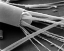
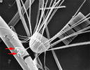
Corethron
|
|
Comments
|
arrows indicate claw spines
|
|
Creator
|
Frank E. Round
|
|
Acknowledgements
|
This image is derived from the Professor Frank Round Image Archive at the Royal Botanic Garden Edinburgh
|
|
Specimen Condition
|
Dead Specimen
|
|
Identified By
|
Frank E. Round
|
|
Life Cycle Stage
|
Vegetative phase
|
|
Body Part
|
valve and spines
|
|
View
|
external, SEM
|
|
Image Use
|
 This media file is licensed under the Creative Commons Attribution-NonCommercial License - Version 3.0. This media file is licensed under the Creative Commons Attribution-NonCommercial License - Version 3.0.
|
|
Copyright
|
© 2008 David G. Mann

|
|
Attached to Group
|
Corethron: view page image collection
|
|
Title
|
corethron2.jpg
|
|
Image Type
|
Photograph
|
|
Image Content
|
Body Parts, Ultrastructure
|
|
ALT Text
|
Corethron spines
|
|
ID
|
29864
|
|
| 29865 |
|
|
Scientific Name
|
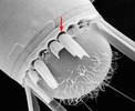
Corethron
|
|
Comments
|
arrow indicates socket
|
|
Creator
|
Frank E. Round
|
|
Acknowledgements
|
This image is derived from the Professor Frank Round Image Archive at the Royal Botanic Garden Edinburgh
|
|
Specimen Condition
|
Dead Specimen
|
|
Life Cycle Stage
|
Vegetative phase
|
|
Body Part
|
theca
|
|
View
|
external detail, showing spines and sockets: SEM
|
|
Image Use
|
 This media file is licensed under the Creative Commons Attribution-NonCommercial License - Version 3.0. This media file is licensed under the Creative Commons Attribution-NonCommercial License - Version 3.0.
|
|
Copyright
|
© 2008 David G. Mann

|
|
Attached to Group
|
Corethron: view page image collection
|
|
Title
|
corethron_sockets.jpg
|
|
Image Type
|
Photograph
|
|
Image Content
|
Body Parts, Ultrastructure
|
|
ALT Text
|
Corethron sockets
|
|
ID
|
29865
|
|
| 29714 |

|
|
| 29713 |
|
|
Scientific Name
|
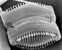
Dimeregramma
|
|
Creator
|
Frank E. Round
|
|
Acknowledgements
|
This image is derived from the Professor Frank Round Image Archive at the Royal Botanic Garden Edinburgh
|
|
Specimen Condition
|
Dead Specimen
|
|
Identified By
|
David Mann
|
|
Life Cycle Stage
|
Vegetative phase
|
|
Body Part
|
Dividing frustule
|
|
View
|
Exterior, SEM
|
|
Image Use
|
 This media file is licensed under the Creative Commons Attribution-NonCommercial License - Version 3.0. This media file is licensed under the Creative Commons Attribution-NonCommercial License - Version 3.0.
|
|
Copyright
|
© 2008 David G. Mann

|
|
Attached to Group
|
Bacillariophyceae: view page image collection
Diatoms: view page image collection
|
|
Title
|
dimeregramma_frustule.jpg
|
|
Image Type
|
Photograph
|
|
Image Content
|
Specimen(s), Ultrastructure
|
|
ALT Text
|
Dimeregramma: dividing frustule
|
|
ID
|
29713
|
|
| 29555 |

|
|
Scientific Name
|
Coscinodiscus
|
|
Location
|
Cultured cells from North Sea marine plankton
|
|
Comments
|
These cells settled into a regular array as a result of slight agitation and vibration of the Petri dish containing them
|
|
Specimen Condition
|
Live Specimen
|
|
Identified By
|
David Mann
|
|
Life Cycle Stage
|
vegetative
|
|
Image Use
|
 This media file is licensed under the Creative Commons Attribution-NonCommercial License - Version 3.0. This media file is licensed under the Creative Commons Attribution-NonCommercial License - Version 3.0.
|
|
Copyright
|
© 2008 David G. Mann

|
|
Creation Date
|
1990s
|
|
Attached to Group
|
Diatoms: view page image collection
|
|
Title
|
coscinodiscusarray.jpg
|
|
Image Type
|
Photograph
|
|
Image Content
|
Specimen(s)
|
|
ALT Text
|
Coscinodiscus
|
|
ID
|
29555
|
|
| 29556 |

|
|
Scientific Name
|
Stephanodiscus
|
|
Creator
|
Frank E. Round
|
|
Acknowledgements
|
This image is derived from the Professor Frank Round Image Archive at the Royal Botanic Garden Edinburgh
|
|
Specimen Condition
|
Dead Specimen
|
|
Identified By
|
David Mann
|
|
Life Cycle Stage
|
Vegetative
|
|
Body Part
|
Valve
|
|
View
|
Exterior, tilted: SEM image
|
|
Image Use
|
 This media file is licensed under the Creative Commons Attribution-NonCommercial License - Version 3.0. This media file is licensed under the Creative Commons Attribution-NonCommercial License - Version 3.0.
|
|
Copyright
|
© David G. Mann

|
|
Title
|
stephanodiscus.jpg
|
|
Image Type
|
Photograph
|
|
Image Content
|
Specimen(s)
|
|
ID
|
29556
|
|
| 29557 |
|
|
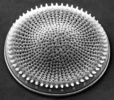
Scientific Name
|
Stephanodiscus
|
|
Creator
|
Frank E. Round
|
|
Acknowledgements
|
This image is from the Professor Frank Round Image Archive at the Royal Botanic Garden Edinburgh
|
|
Specimen Condition
|
Dead Specimen
|
|
Identified By
|
David Mann
|
|
Life Cycle Stage
|
Vegetative phase
|
|
Body Part
|
Valve
|
|
View
|
Exterior, tilted
|
|
Image Use
|
 This media file is licensed under the Creative Commons Attribution-NonCommercial License - Version 3.0. This media file is licensed under the Creative Commons Attribution-NonCommercial License - Version 3.0.
|
|
Copyright
|
© 2008 David G. Mann

|
|
Title
|
stephanodiscus1.jpg
|
|
Image Type
|
Photograph
|
|
Image Content
|
Specimen(s)
|
|
Technical Information
|
SEM image
|
|
ALT Text
|
Stephanodiscus valve
|
|
ID
|
29557
|
|
| 29558 |

|
|
Scientific Name
|
Hydrosera
|
|
Creator
|
Frank E. Round
|
|
Acknowledgements
|
This image is derived from the Professor Frank Round Image Archive at the Royal Botanic Garden Edinburgh
|
|
Specimen Condition
|
Dead Specimen
|
|
Identified By
|
David Mann
|
|
Life Cycle Stage
|
Vegetative phase
|
|
Body Part
|
Whole frustule, SEM
|
|
View
|
Exterior
|
|
Image Use
|
 This media file is licensed under the Creative Commons Attribution-NonCommercial License - Version 3.0. This media file is licensed under the Creative Commons Attribution-NonCommercial License - Version 3.0.
|
|
Copyright
|
© David G. Mann

|
|
Attached to Group
|
Diatoms: view page image collection
|
|
Title
|
hydrosera.jpg
|
|
Image Type
|
Photograph
|
|
Image Content
|
Specimen(s)
|
|
Technical Information
|
Scanning electron micrograph
|
|
ALT Text
|
Hydrosera frustule
|
|
ID
|
29558
|
|
| 29559 |
|
|
Creator
|
Frank E. Round
|
|
Acknowledgements
|
This image is derived from the Professor Frank Round Image Archive at the Royal Botanic Garden Edinburgh
|
|
Specimen Condition
|
Dead Specimen
|
|
Identified By
|
David Mann
|
|
Life Cycle Stage
|
Vegetative phase
|
|
Body Part
|
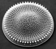
Valve
|
|
View
|
External view, SEM
|
|
Image Use
|
 This media file is licensed under the Creative Commons Attribution-NonCommercial License - Version 3.0. This media file is licensed under the Creative Commons Attribution-NonCommercial License - Version 3.0.
|
|
Copyright
|
© David G. Mann

|
|
Attached to Group
|
Stephanodiscus (Thalassiosirales): view page image collection
|
|
Title
|
stephanodiscus2.jpg
|
|
Image Type
|
Photograph
|
|
Image Content
|
Specimen(s)
|
|
Technical Information
|
Scanning electron micrograph
|
|
ALT Text
|
Stephanodiscus valve
|
|
ID
|
29559
|
|
Please note: Most images and other media displayed on the Tree of Life web site are protected by copyright, and the ToL cannot act as an agent for their distribution. If you would like to use any of these materials for your own projects, you need to ask the copyright owner(s) for permission. For additional information, please refer to the
ToL Copyright Policies.


 This media file is licensed under the
 Go to quick links
Go to quick search
Go to navigation for this section of the ToL site
Go to detailed links for the ToL site