


Abralia multihamata
Kotaro Tsuchiya and Richard E. Young

Introduction
A. multihamata is a species endemic to the shelf waters along the south of Sagami Bay, Japan to Taiwan and off Papua New Guinea. It is characterized by the scattered arrangement of ventral mantle photophores, six club hooks, and more than five eye photophores of two types. The taxonomic status with A. spaercki is problematic.
Sasaki's specimens of A. multihamata ranged from 25-32 mm ML. Voss (1963) described A. lucens from specimens that ranged from 43-62 mm ML. The title illustrations show a 35 mm ML A. multihamata and a 62 mm ML A. lucens. The two look very different. Tsuchiya and Okutani (1988) suggest this difference is due to growth and A. lucens is included here as a junior synonym of A. multihamata. This status, however, needs confirmation.
Brief diagnosis:
An Abralia (Abralia) without ...
- peculiar photophore in the funnel groove.
- scattered photophores between medial and lateral arm photophores on arms IV.
Characteristics
- Tentacle club
- About six hooks on ventral side.
- Two rows of large suckers on dorsal side.
Click on an image to view larger version & data in a new window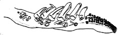
Figure. Oral view of the tentacular club of A. multihamata, Sagami Bay. Drawing from Tsuchiya (2000).
- Hectocotylus
- Right ventral arm of male hectocotylized.
- Hectocotylus with two different-sized offset flaps.
Click on an image to view larger version & data in a new window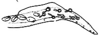
Figure. Oral view of the distal portion of the hectocotylus of A. multihamata, Sagami Bay. Drawing from Tsuchiya (2000).
- Eye Photophores
- Five major organs: two large opaque, terminal organs and three intermediate silvery organs; small silvery additional organs present.
Click on an image to view larger version & data in a new window

Figure. Ventrolateral view of the ocular photophores of A. multihamata. Left - male, Japanese waters. Drawing from Sasaki (1929). Right - Sex ?, Solomon Sea off South New Britain. Photograph by Jeffrey Dubosc, SPC.
- Integument Photophores
- Ventral head with indistinct striped arrangement of integumental organs.
- Ventral arms without photophores between medial and lateral arm photophore series.
Click on an image to view larger version & data in a new window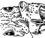
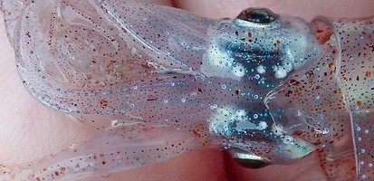
Figure. Ventral view of the head photophores of A. multihamata. Left - male, Japanese waters.Drawing from Sasaki (1929). Right - Sex ?, Solomon Sea off South New Britain. Photograph by Jeffrey Dubosc, SPC.
- Ventral mantle with photophores with scattered arrangement of integumental organs but with distinct bare stripe in midline. Click on an image to view larger version & data in a new window
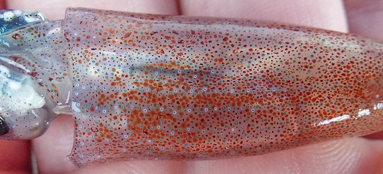
Figure. Ventral view of the head photophores of A. multihamata. sex ?, Solomon Sea off South New Britain. The two distinct rows of lensed photophores define the midline bare stripe. Photograph by Jeffrey Dubosc, SPC.
- Reproductive structures
- Spermatangia receptacles. Receptacles are located underneath the dorsal collar on either side of the nuchal cartilage. Pigment marking the receptacles is visible through the muscular walls of the collar. Within the collar pocket is an inner muscular (?) lid that carries the pigment seen externally. The spermatangia seem to attach beneath the inner lid where it joins the lateral wall of the nuchal cartilage.
Click on an image to view larger version & data in a new window
Figure. Spermatangia receptacles of A. multihamata. Left - Dorsal view with the mantle reflected posteriorly showing the collars on either side of the dorsal rim of the nuchal cartilage. The white arrow points to the pigmented inner lid of one of the two receptacles. Right - Dorsolateral view with one collar cut and its dorsal portion folded upward (and impaled by a dissecting pin) revealing the edge of the inner lid and the spermatangia beneath it which seem to be encased in a mucous mass.
- Spermatangia receptacles. Receptacles are located underneath the dorsal collar on either side of the nuchal cartilage. Pigment marking the receptacles is visible through the muscular walls of the collar. Within the collar pocket is an inner muscular (?) lid that carries the pigment seen externally. The spermatangia seem to attach beneath the inner lid where it joins the lateral wall of the nuchal cartilage.
Comments.
A. multihamata is very similiar to A. spaercki but lacks the scattered photophores in the proximal half of each arm IV found between the medial and lateral (not lateral membrane) photophore series and lacks the single, large white photophore found in the funnel groove posteromedial to each first occipital lobe in A. spaercki.
Life history
Spent specimens are collected in large number from the East China Sea in October by bottom trawl.Distribution
Vertical distribution
A. multihamata is possibly bottom associated or mesopelagic boundary species. Specimens were collected by bottom trawl from the East China Sea.
Geographical distribution
This species was first described from Taiwan waters. It is distributed in shelf waters of the East China Sea, northward to Sagami Bay and the eastern sector of the Japan Sea. It is also found in the South Pacific off Papua New Guinea.
References
Sasaki, M. 1929. A Monograph of the Dibranchiate Cephalopods of the Japanese and Adjacent Waters. Journal of the College of Agriculture, Hokkaido Imperial University,357 pp.
Tsuchiya, K. 2000. Illustrated book of the Enoploteuthidae. In: Okutani T., ed. True face of Watasenia scintillans. Tokai University Press, Tokyo, p 196269. (in Japanese)
Tsuchiya, K. and T. Okutani. 1988. Subgenera of Enoploteuthis, Abralia and Abraliopsis of the squid family Enoploteuthidae (Cephalopoda, Oegopsida). Bulletin of the National Science Museum, Tokyo (series A), 14: 119-136.
Voss, G.L. 1963. Cephalopods of the Philippine Islands. United States National Museum Bulletin, 234:1-180.
Title Illustrations

| Scientific Name | Abralia multihamata |
|---|---|
| Location | Sagami Bay, Japan |
| Reference | Sasaki, M. 1929. A Monograph of the Dibranchiate Cephalopods of the Japanese and Adjacent Waters. Journal of the College of Agriculture, Hokkaido Imperial University,357 pp. |
| Sex | Male |
| Life Cycle Stage | Mature |
| View | Ventral |
| Size | 35 mm ML |
| Scientific Name | Abralia lucens =? A. multihamata |
|---|---|
| Location | Philippine waters |
| Creator | N. Voss via G. Voss |
| Acknowledgements | Voss, G.L. 1963. Cephalopods of the Philippine Islands. United States National Museum Bulletin, 234:1-180. |
| Sex | Female |
| View | Ventral |
| Size | 62 mm ML |
| Type | Holotype |
About This Page

Tokyo University of Fisheries, Tokyo, Japan
University of Hawaii, Honolulu, HI, USA
Correspondence regarding this page should be directed to Richard E. Young at
Page copyright © 2018 and
 Page: Tree of Life
Abralia multihamata .
Authored by
Kotaro Tsuchiya and Richard E. Young.
The TEXT of this page is licensed under the
Creative Commons Attribution-NonCommercial License - Version 3.0. Note that images and other media
featured on this page are each governed by their own license, and they may or may not be available
for reuse. Click on an image or a media link to access the media data window, which provides the
relevant licensing information. For the general terms and conditions of ToL material reuse and
redistribution, please see the Tree of Life Copyright
Policies.
Page: Tree of Life
Abralia multihamata .
Authored by
Kotaro Tsuchiya and Richard E. Young.
The TEXT of this page is licensed under the
Creative Commons Attribution-NonCommercial License - Version 3.0. Note that images and other media
featured on this page are each governed by their own license, and they may or may not be available
for reuse. Click on an image or a media link to access the media data window, which provides the
relevant licensing information. For the general terms and conditions of ToL material reuse and
redistribution, please see the Tree of Life Copyright
Policies.
- Content changed 11 October 2015
Citing this page:
Tsuchiya, Kotaro and Richard E. Young. 2015. Abralia multihamata . Version 11 October 2015 (under construction). http://tolweb.org/Abralia_multihamata/19658/2015.10.11 in The Tree of Life Web Project, http://tolweb.org/


