


Abraliopsis
Richard E. Young and Kotaro Tsuchiya
- Abraliopsis (Abraliopsis)
- Abraliopsis (Boreabraliopsis) felis
- Abraliopsis (Micrabralia)
- Abraliopsis (Pfefferiteuthis)
Introduction
Species of Abraliopsis are small squids that are most easily recognized by spherical "black" photophores (usually three) at the tip of each Arm IV. The black appearance is due to black chromatophores that tightly cover the photophores but when the latter luminesce, the chromatophores withdraw from the photophores. This feature is shared with Watasenia scintillans from waters around Japan.
Brief diagnosis:
An enoploteuthid ...
- with 2 or 3large, black photophores at the tips of arms IV.
- with hooks of manus in two series.
- with darkly pigmented buccal membrane (pigment not in chromatophores).
Characteristics
- Arms
- Suckers at arm tips in two series; suckers absent from arms IV (only hooks present).
- Right arm IV hectocotylized.
- Attachment of buccal connectives to arms: DDVD.
- Tentacles
- Manus of club with two series of hooks and one series, usually incomplete, of suckers.
Click on an image to view larger version & data in a new window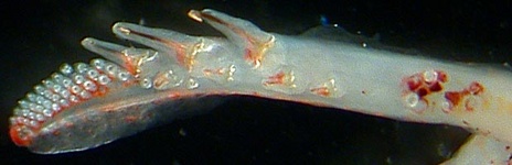
Figure. Oral view of the club of Abraliopsis (Abraliopsis) morisii.
- Manus of club with two series of hooks and one series, usually incomplete, of suckers.
- Buccal crown
- Buccal crown with dark epithelial pigmentation on oral surface rather than typical chromatophores.*
- Buccal connectives connect to ventral borders of arms III. Note: some species of Abralia differ in this character.
Click on an image to view larger version & data in a new window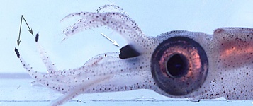
Figure. Side view of Abraliopsis sp. B. White arrow points to pigmented buccal crown. Black arrows point to large, pigmented photophores at the tips of arms IV. Photograph by R. Young.
- Fins
- Fins extend to posterior end of tail.
- Fins extend to posterior end of tail.
- Photophores. As in Abralia and Watasenia, Abraliopsis has three types of common integumental photophores covering the arms, head, funnel and mantle. In the photos above the most complex of the integumental photophores with red color filters (called "complex" or "red" photophores) are easily recognized. The other two types of common integumental photophores (called "non-complex or "blue" photophores) have a blue color. The latter exhibit a variety of sizes and the largest of these, the second type, are photophores bearing large lenses. The smaller ones are the third type which are lensless. A fourth, but uncommon type of integumental photophore, is the colorless (ie, "white") photophores known only from the funnel groove. The structure of this type has not been investigated but these few photophores are clearly not all of the same structure.
- Two to four large photophores covered by black chromatophores on tips of each arm IV (see photograph under "Buccal crown.")*
- Five photophores on eyeball.*
- Complex photophores of integument, in life, with red-colored filters.*
- Photophores of funnel groove on head, distinctly different with white color.*
Click on an image to view larger version & data in a new window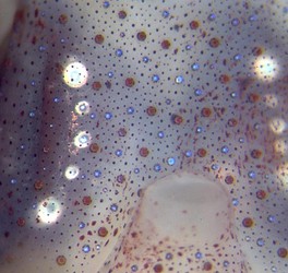
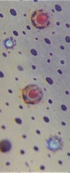
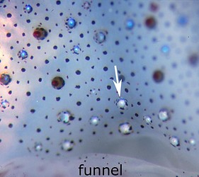
Figure. Ventral surface of the head and photophores of Abraliopsis sp. B. Left - Three types of photophores are apparent: Small photophores with red filters, smaller photophores with a blue reflection and large photophores (the ocular photophores) beneath the skin on the ventral surfaces of the eyeballs. Middle - An enlargement of some of the skin photophores. Right - white photophores of funnel groove (hidden by the funnel in the middle photograph). The arrow points to the anterior-most white photophore in the midline of the funnel groove. To the side and above the arrow are the "red" and "blue" photophores. Photographs by R. Young.
*Characters shared with Watasenia.
- Photophore terminology: Terminology for photophore distribution patterns can be found here.
- Spermatangia receptacles
- Located under dorsal collar or under gladius between stellate ganglia or both sites.
- Located under dorsal collar or under gladius between stellate ganglia or both sites.
Classification
Species of Abraliopsis can be placed into four subgenera but the subgenus Micrabralia is probably not a natural group. The following table shows the primary taxonomic features of each group. See a more detailed comparison A. (Boreabraliopsis) and A. (Abraliopsis) on the A. (B.) felis page.
| Abraliopsis (Boreabraliopsis) | Abraliopsis (Abraliopsis) | Abraliopsis (Micrabralia) | Abraliopsis (Pfefferiteuthis) | |
| No. of species | 1 | 7 | 5 | 7 |
| Oral surface of arms in males | Without papillae | Without papillae | With or without some papillae | Some or all arms with papillae |
| Ventral protective membrane of left arm IV, male, | Virtually absent | Virtually absent | Variable but not greatly enlarged | Greatly enlarged membrane and trabeculae, with papillae |
| Photophore arrangement on ventral surface of head | Scattered | Scattered | Red and blue integumental photophores in linear pattern, with or without additional scattered blue photophores. | Red and blue integumental photophores in linear pattern, with or without additional scattered blue photophores. |
| Length of dorsal flap of hectocotylus relative to ventral flap | Nearly equal | Absent to very small (~ third) | ~ third to half | ~ half |
| Pattern* of funnel-groove photophores. | Simple Pattern: | Moderate Pattern:  | Moderate (top) or Complex-1 Pattern (bottom):  | Complex-2 Pattern: |
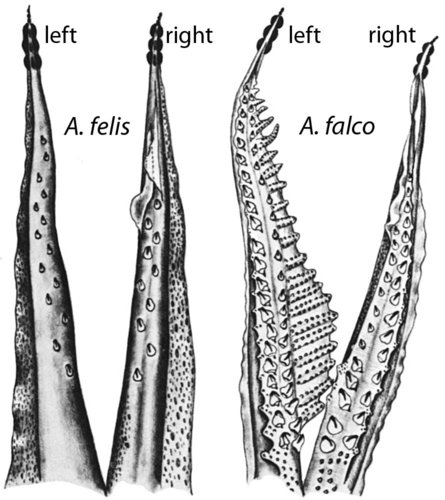
Figure. Arms IV from arms IV showing some of the group characters. Note the papillae at the base of the arms in A. falco and the smooth arms in A. felis, and the greatly expanded ventral membrane on the left arm IV of A. falco. Drawings from Young (1972).
References
Tsuchiya, K. and T. Okutani. 1988. Subgenera of Enoploteuthis, Abralia and Abraliopsis of the squid family Enoploteuthidae (Cephalopoda, Oegopsida). Bulletin of the National Science Museum, Tokyo (series A) 14: 119-136.
Young, R.E. 1972. The systematics and areal distribution of pelagic cephalopods from the sea off southern California. Smiths. Contr. Zool., 97:1-159.
Young, R. E., L. A. Burgess, C. F. E. Roper, M. J. Sweeney and S. J. Stephen. 1998. Classification of the Enoploteuthidae, Pyroteuthidae and Ancistrocheiridae. Smithson. Contr. Zool., No. 586: 239-256.
Title Illustrations

| Scientific Name |
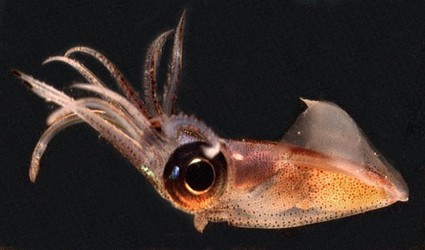
Abraliopsis sp. B Abraliopsis |
|---|---|
| Location | Hawaiian waters |
| Specimen Condition | Live Specimen |
| View | Side |
| Image Use |
 This media file is licensed under the Creative Commons Attribution-NonCommercial License - Version 3.0. This media file is licensed under the Creative Commons Attribution-NonCommercial License - Version 3.0.
|
| Copyright |
© 2000

|
About This Page
University of Hawaii, Honolulu, HI, USA
Tokyo University of Fisheries, Tokyo, Japan
Page copyright © 2013 and
 Page: Tree of Life
Abraliopsis .
Authored by
Richard E. Young and Kotaro Tsuchiya.
The TEXT of this page is licensed under the
Creative Commons Attribution-NonCommercial License - Version 3.0. Note that images and other media
featured on this page are each governed by their own license, and they may or may not be available
for reuse. Click on an image or a media link to access the media data window, which provides the
relevant licensing information. For the general terms and conditions of ToL material reuse and
redistribution, please see the Tree of Life Copyright
Policies.
Page: Tree of Life
Abraliopsis .
Authored by
Richard E. Young and Kotaro Tsuchiya.
The TEXT of this page is licensed under the
Creative Commons Attribution-NonCommercial License - Version 3.0. Note that images and other media
featured on this page are each governed by their own license, and they may or may not be available
for reuse. Click on an image or a media link to access the media data window, which provides the
relevant licensing information. For the general terms and conditions of ToL material reuse and
redistribution, please see the Tree of Life Copyright
Policies.
- Content changed 03 November 2013
Citing this page:
Young, Richard E. and Kotaro Tsuchiya. 2013. Abraliopsis . Version 03 November 2013 (under construction). http://tolweb.org/Abraliopsis/19644/2013.11.03 in The Tree of Life Web Project, http://tolweb.org/


