


Asperoteuthis mangoldae
Richard E. Young, Michael Vecchione, and Clyde F. E. Roper
Introduction
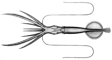
Figure. Asperoteuthis mangoldae, holotype, 80 mm ML. Drawing by A. Hart.
Asperoteuthis mangoldae (first known as Asperoteuthis sp. A on this page) is very gelatinous, with about the same consistency as Grimalditeuthis bonplandi. It has elongate ventral arms in small squid, but these get relatively short in large squid. It lacks the tubercles and elongate fins of A. acanthoderma.
Brief diagnosis:
An Asperoteuthis ...- without integumental tubercules.
- with comma-shaped groove in funnel locking-apparatus.
Characteristics
- Arms
- Largest arm suckers with 6-10 truncated teeth on distal half of ring.
- Arms without enlarged suckers; largest suckers in proximal half of arm with sucker size slowly decreasing distally; arms I-III with suckers of comparable size, arms IV with distinctively smaller suckers.
- Tentacles
- Suckers with ca. 8 large, truncated teeth on distal half of inner ring grading to ca. 17 small, truncated teeth on proximal half.
- Club with very broad proximal protective membrane and long terminal papilla.
Click on an image to view larger version & data in a new window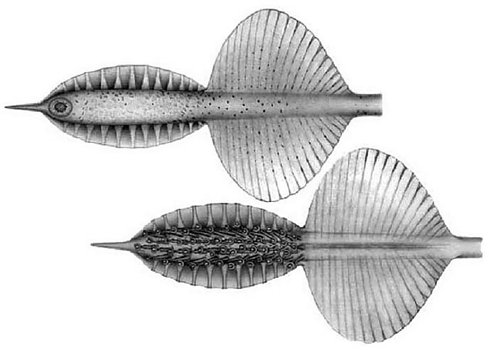
Figure. Aboral (top) and oral (bottom) views of the tentacular club of A. mangoldae, NMNH 729749, holotype. Drawing by A. Hart.
- Head
- Beaks: Descriptions can be found here: Lower beak; upper beak.
- Beaks: Descriptions can be found here: Lower beak; upper beak.
- Funnel
- funnel-locking apparatus oval with a rounded ridge in the midline (tragus?) and an elongate, deep, curving groove laterally; mantle locking component with matching curved ridge. Antitragus absent.
Click on an image to view larger version & data in a new window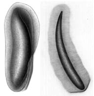
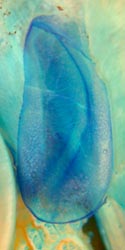
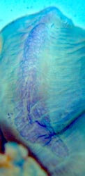
Figure. Frontal views of the funnel-mantle locking apparatus of A. mangoldae. Left - Funnel and mantle components (left side), NMNH 729752. Drawings by A. Hart. Right - Funnel and mantle components (left side), 100 mm ML, mature male, paratype, stained with methylene blue. Photographs by R. Young.
- funnel-locking apparatus oval with a rounded ridge in the midline (tragus?) and an elongate, deep, curving groove laterally; mantle locking component with matching curved ridge. Antitragus absent.
- Mantle
- Mantle and other integument without tubercles.
- Mantle and other integument without tubercles.
- Fins
- Fin length 40% of ML.
- Fin width approximately equals fin length.
- Photophores
- Club-tip photophore small, terminal papilla large.
- Small embedded photophores present on aboral surface of club.
- Ventral eyeball photophore present.
- Luminescent pads present on tentacle stalks.
Comments
More details of the description can be found here.
A. mangoldae apparently possesses a large secondary tail but a specimen with an attached tail has not been captured.
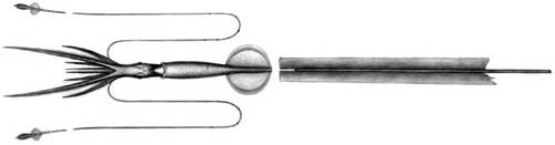
Figure. The drawing is a reconstruction based on the collection of a damaged tail in the same tow with a small (80 mm ML) A. mangoldae. The tail did not match the broken end of the gladius from the specimen indicating that a piece of the tail was missing. Tentacular clubs, missing from specimen, were added to drawing. Drawing by A. Hart.
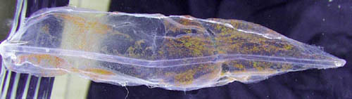
Figure. The photograph shows the tail of a large Asperoteuthis from the same locality and possibly belonging to a large A. mangoldae although the shape differs greatly from that of the small specimen above.
Very few A. mangoldae have been captured with the clubs attached as the long, thin tentacles are easily broken off during capture. With tentacles missing, it can be confused with Grimalditeuthis bomplandi in its general shape and consistency. However, many differences easily separate them including presence of the funnel-mantle fusion and three large papillae on each arm-sucker base in G. bomplandi.
In addition to the features listed in the Diagnosis, A. mangoldae differs from A. acanthoderma in (1) the weaker consistency of the arms and mantle, (2) the dentition of the arm (6-10 vs 3-4) and club (25 vs 9 teeth) suckers, (3) the smaller and more circular terminal club photophore, (4) the longer terminal papilla on the club and (5) the broader fins (width ca 115% of length vs ca 75% of length). In addition, the club suckers of A. acanthoderma are on shorter stalks, the sucker rings are more elongated in the oral-aboal directiion and suckers of arms IV are more densely packed.
Behavior
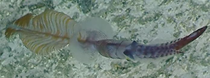
This squid was observed swimming slowly above the ocean floor at 930 m depth on Jarvis Seamount, from an ROV operated by the E/V (Exploration Vessel) Nautilus. When first seen (i.e., the title image on this page) it had a, broad, striped leaf-shaped tail (or more accurately a tail that looks like a sea pen as one shipboard observer noted) posterior to the fins. About 10 seconds later it changed direction and moved more quickly toward the ROV while collapsing the leaf-shape. To the great surprise of the observers and the authors of this page, within about 5 seconds the leaf-shape had almost disappeared, held tightly onto the extended rod-like gladius of the tail. The squid now looked like the squid below although this photograph was taken about a minute later.
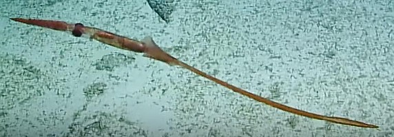
Figure. Asperoteuthis cf mangoldae, ROV image taken at a depth of 930 m. Note that the "secondary fins" have collasped onto the tail. © 2019 Ocean Exploration Trust/Nautilus Live
As the squid started to move toward the ROV while its leaf-shaped tail collapsed, its arms flared outward momentarily but returned to their normal posture as it disappeared off the bottom of the screen. Moments later it reappeared, swimming tail first and quickly moving away. When the operator of the ROV steared to follow the squid, it, immediately, reversed direction, flared arms and tentacles, then folded them under the head, as it rapidly swam toward the ROV head-first. One observer stated, "This one looks like it is trying to fight us" as the squid swam off the bottom of the screen. It soon reappeared moving slowly tail first with arms and tentacles flared, then it squirted a jet of ink while retracting arms and tentacles and it swam rapidly away. The ROV operator was able to find the squid again resulting in the frame grab above this paragraph. You can see the video here.
The squid was identified by the distinctive form of the expanded tail as belonging to Asperoteuthis mangoldae. As indicated on this page the occurrence of this type of tail in this species is likely, but it is not yet proven. Because the squid was not captured the identification cannot be confirmed.
Life History
Paralarvae of A. mangoldae from off Hawaii have been described by Young, 1991. The young stages are very similar to those of Chiroteuthis picteti in the virtual lack of a brachial pillar and the centrally located esophagus. It differs, however, by:
- a chromatophore on the oral surface of each tentacle base
- the greater number of arm suckers
- more numerous dorsal brachial pillar vesicles
- the lack of a groove in the funnel cartilage
- the dorsally elongate vesiculate area on the mantle (at 6 mm ML)
At larger sizes the triangular shape of the dorsal-mantle, vesiculate area, the more circular shape of the combined fins and the straight funnel locking-cartilage are diagnostic. In the large paralarvae (i.e., ca. 27 mm ML) the adult club begins to appear on the tentacle stalk and the bare proximal region is denoted by the broader protective membrane in this area.
In larger paralarvae the elongate vesiculate area on the dorsal mantle is the diagnostic feature most easliy recognized.
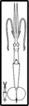
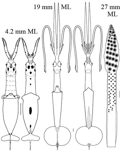
Figure. A. mangoldae paralarvae. Thumbnail (far left) - Ventral views showing relative sizes of the two paralarvae. Left - Ventral and dorsal views, 4.2 mm ML. Middle - Ventral and dorsal views, 19 mm ML. Right - Oral view of the tentacular club, 27 mm ML, with a typical primary doratopsid club (the distal expanded region with large suckers). The developing adult club is seen proximal to the doratopsid club on the tentacular stalk. This consists of a distal region with virtually no protective membranes and small suckers, and a proximal region with narrow protective membranes and no suckers. The developing adult club, therefore, shows the beginning of the features characteristic of the subadult. Scale bars = 1mm. Drawings from Young (1991).
Distribution
Vertical distribution
Off Hawaii eight specimens were captured during a study to examine the vertical distribution of cephalopods. All specimens were captured during the day at depths between 775 and 975 m. The absence of captures in the heavily sampled waters from 0-400m depth at night suggests that this species is not a vertical migrator.
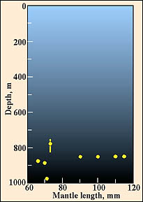
Figure. Vertical distribution chart of A. mangoldae. Captures were made with both open and opening/closing trawls. Bars - fishing depth-range of opening/closing trawl. Circle - Modal fishing depth for either trawl. Yellow-filled circles - Day capture.
Geographical distribution
Type locality: Hawaiian waters at 21°25'N, 158°20.5'W. The holotype was captured in an opening-closing trawl between 820 and 870 m depth during the day (1411-1616 hrs). This species is known only from the waters off the Hawaiian Islands. Both A. mangoldae and Asperoteuthis acanthoderma occur in these waters but A. mangoldae is much more abundant.
References
Young, R. E. 1978. Vertical distribution and photosensitive vesicles of pelagic cephalopods from Hawaiian waters. Fish. Bull., 76: 583-615.
Young, R. E., M. Vecchione and C. F. E. Roper. 2007. A new genus and three new species of decapodiform cephalopods (Mollusca: Cephalopoda). Rev. Fish. Biol. Fisheries, 17: 353-365.
Title Illustrations

| Scientific Name | Asperoteuthis mangoldae |
|---|---|
| Location | Jarvis Seamount, Equatorial Pacific at 0°23'S, 159°59'W, depth 930 m. |
| Comments | Photographed from the ROV Hercules |
| Acknowledgements | Officers and crew of the Exploration Vessel (E/V) Nautilus |
| Specimen Condition | Live Specimen |
| Identified By | Michael Vecchione |
| Behavior | "Secondary fins" of tail expanded. |
| View | Dorsal |
| Copyright | © 2019 Ocean Exploration Trust/Nautilus Live |
About This Page

University of Hawaii, Honolulu, HI, USA
National Museum of Natural History, Washington, D. C. , USA
Smithsonian Institution, Washington, D. C., USA
Page copyright © 2019 , , and
 Page: Tree of Life
Asperoteuthis mangoldae .
Authored by
Richard E. Young, Michael Vecchione, and Clyde F. E. Roper.
The TEXT of this page is licensed under the
Creative Commons Attribution-NonCommercial License - Version 3.0. Note that images and other media
featured on this page are each governed by their own license, and they may or may not be available
for reuse. Click on an image or a media link to access the media data window, which provides the
relevant licensing information. For the general terms and conditions of ToL material reuse and
redistribution, please see the Tree of Life Copyright
Policies.
Page: Tree of Life
Asperoteuthis mangoldae .
Authored by
Richard E. Young, Michael Vecchione, and Clyde F. E. Roper.
The TEXT of this page is licensed under the
Creative Commons Attribution-NonCommercial License - Version 3.0. Note that images and other media
featured on this page are each governed by their own license, and they may or may not be available
for reuse. Click on an image or a media link to access the media data window, which provides the
relevant licensing information. For the general terms and conditions of ToL material reuse and
redistribution, please see the Tree of Life Copyright
Policies.
- Content changed 20 August 2019
Citing this page:
Young, Richard E., Michael Vecchione, and Clyde F. E. Roper. 2019. Asperoteuthis mangoldae . Version 20 August 2019 (under construction). http://tolweb.org/Asperoteuthis_mangoldae/19467/2019.08.20 in The Tree of Life Web Project, http://tolweb.org/


