


Grimalditeuthis
Grimalditeuthis bonplandi
Richard E. Young and Clyde F. E. Roper

Introduction
Grimalditeuthis bonplandi is very gelatinous and contains vesiculate tissue in the head, arms and mantle. It is generally Chiroteuthis-like in appearance (long neck, body and fin shape) but is distinguished by subequal arms and fusion of each funnel-mantle locking apparatus. Only scattered chromatophores are present, and the eyes are small. This species is infrequently captured but seems to have a world-wide distribution in tropical to temperate seas.
Brief diagnosis:
A chiroteuthid ...
- without suckers on clubs.
- with funnel fused to mantle at each funnel-mantle lock.
- without photophores except on arm tips in mature (?) females.
Characteristics
- Arms
- Arms approximately subequal in length, gelatinous.
- Sucker base with three conical papillae (unique character).
- Protective membranes absent. Click on an image to view larger version & data in a new window
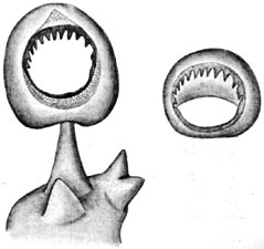

Figure. Oral view of large arm III sucker, stalk and base, with inner ring at right, G. bonplandi, 89 mm ML, female, off Southern California. Drawing from Young (1972).
- Tentacles
- Club divided into two portions by symmetrical protective membranes.
- Suckers absent from clubs. Click on an image to view larger version & data in a new window
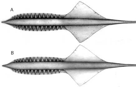
Figure. Tentacle-clubs of G. bomplandi. A - Aboral view. B - Oral view. a single intact tentacle was found on a specimen taken from the stomach of the fish Alepisaurus ferox (courtesy of Lourdes Burgess). The tentacle club lacked suckers and showed no indication that suckers or sucker stalks were ever present as the skin was intact. Drawings by A. D. Hart.
- Head
- Olfactory organ located lateral to base of funnel (i.e., immediately anterior to collar at posterior end of neck).
- Beaks: Descriptions can be found here: Lower beak; upper beak.
- Olfactory organ located lateral to base of funnel (i.e., immediately anterior to collar at posterior end of neck).
- Funnel
- Funnel valve present.
- Funnel fused to mantle at each funnel-mantle locking-apparatus (head not fused to mantle in nuchal region).
- Photophores
- Absent accept at the arm tips of mature (?) females (See "More details ...").
- Absent accept at the arm tips of mature (?) females (See "More details ...").
- Tail
- Tail retained in adults with two fin-like "floatation devices" (= secondary fins) arise from tail.
Comments
More details of the description can be found here.The function of the suckerless club is unknown. The extremely thin tentacle stalks (these fragile structures are almost always lost during capture by trawls) indicate that typical tentacle deployment would be difficult. The proximal fused trabeculae of the club can function as fins that can flap to extend the tentacle. This combined with wiggling of the muscular core of the club indicates that the club can swim itself into position, and once in position, wiggling of the club might attract potential prey for the squid (Hoving, et al., 2015). In contrast to extension of the tentacle, retraction can occur quickly as the squid swims towards the club.
Look at this one also.
This species also has a very characteristic pattern of chromatophores on the head. A line of chromatophores passes across the ventral surface of the head between the anterior ends of eyes; another line runs along the neck from each olfactory papilla anteriorly to each eye, then anterior to each eye along the brachial pillar, terminating at the base of the arms.
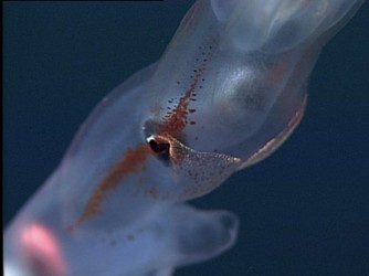
Figure. Insitu ventrolateral view of the head of G. bonplandi showing the characteristic chromatophore strip along the side of the head and a previously unrecognized iridescent patches on the ventral side of the head. Photograph © 2011 MBARI.
Nomenclature
Joubin (1898) described a second species, G. richardi based on the presence of photophores on the tips of the arms. Pfeffer (1912) synonomized the two and this was supported by Young (1972) and Nesis (1982).
Life History
The doratopsis was first described by Chun (1910) as Doratopsis sagitta. A growth series of the paralarvae was described by Young (1991), and can be found below. The early doratopsis stage is similar to that of other species in the family. The older doratopsis shows many of the features of the subadult including the distinctive chromatophore pattern and the small eyes.
- At sizes larger than 9 mm ML Grimalditeuthis doratopsis paralarvae are easily identified by:
- Small eye size.
- Position of the olfactory papillae opposite the base of the funnel.
- Distinctive chromatophore pattern on the head (same as subadult pattern).
- Separation of optic lobes from brain.
- Anterior position of the superior buccal and brachial lobes.
- At sizes smaller than 9 mm paralarvae are identified by a combination of:
- A long brachial pillar with a centrally located esophagus.
- Number of chromatophores on the funnel shoulders and the ventral head posterior to the eyes.
- Just slightly elongate eye-shape.
- Shape of the vesiculate area on the posterior end of the mantle (nearly flat anteriorly).
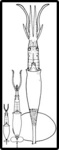

Figure. Paralarvae of G. bonplandi, Hawaiian waters. Thumbnail - Ventral view of the three paralarvae showing relative sizes. A - Side view of the head showing the long brachial pillar and the central position of the esophagus. B-D - Dorsal and ventral views of paralarvae at three different sizes. E - Oral view of the tentacular club. Proximal to the typical doratopsid club there is a scattered arrangement of suckers which contrasts with the denser and more orderly arrangement of presumptive adult clubs in advanced paralarvae of Chiroteuthis and Asperoteuthis. Scale bars are 1 mm. Drawings from Young (1991)
Distribution
Vertical distribution
A small number of captures off Hawaii (Young 1978) included three small squid from the upper 350 m that probably had not descended from the shallow paralarval habitat and five squid from depths greater than 700 m.


Figure. Vertical distribution chart of G. bonplandi, Hawaiian waters. Captures were made with both open and opening/closing trawls. Bars - Fishing depth-range of opening/closing trawl. Circle - Modal fishing depth for either trawl. Blue color - Night captures. Yellow color - Day captures. Chart modified from Young (1978).
Geographical distribution
The type locality is 29° N and 39° W in the North Atlantic. It is known from the tropical and subtropical North Atlantic and the tropical and temperate North Pacific (Nesis, 1982).
References
Hoving, H.J.T., L. D. Zeidberg, M. C. Benfield, S. L. Bush, B. H. Robison and M. Vecchione. 2015. First in situ observations of the deep-sea squid Grimalditeuthis bonplandi reveal unique use of tentacles. Proc. R. Soc. B 280: 20131463. http://dx.doi.org/10.1098/rspb.2013.1463
Joubin, L. 1898. Observations sur divers Cephaloppodes. Quatrieme note: Grimalditeuthis Richardi Joubin 1898. Bulletin de la Societe Zoologique de France, 23: 101-113.
Pfeffer, G. 1912. Die Cephalopoden der Plankton-Expedition. Ergebniss der Plankton-Expedition der Humboldt-Stiftung. 2: 1-815.
Verany, J. B. 1839. Memoire sur six novelles especes de Cephalopodes trouvees dans la Mediterranee a Nice. Memoire della Resle Accademia della Science de Torino, series 2, 1: 91-98
Young, R. E. 1972. The systematics and areal distribution of pelagic cephalopods from the seas off Southern California. Smithson. Contr. Zool., 97: 1-159.
Young, R. E. 1978. Vertical distribution and photosensitive vesicles of pelagic cephalopods from Hawaiian waters. Fish. Bull., 76: 583-615.
Young, R. E. 1991. Chiroteuthid and related paralarvae from Hawaiian waters. Bull. Mar. Sci. 49: 162-185.
Title Illustrations
| Scientific Name | Grimalditeuthis bonplandi |
|---|---|
| Location | Off Southern California |
| Reference | modified from Young, R. E. 1972. The systematics and areal distribution of pelagic cephalopods from the seas off Southern California. Smithson. Contr. Zool. 97:1-159. |
| Sex | Female |
| View | Ventral |
| Size | 89 mm ML |
| Image Use |
 This media file is licensed under the Creative Commons Attribution-NonCommercial License - Version 3.0. This media file is licensed under the Creative Commons Attribution-NonCommercial License - Version 3.0.
|
| Copyright |
©

|

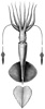
| Scientific Name | Grimalditeuthis bonplandi |
|---|---|
| Location | Hawaiian waters |
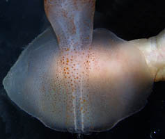
| Comments | The photograph shows the delicate, translucent nature of the fin and its light pigmentation. A human finger can be seen behind the fin. |
| Body Part | Fins |
| View | Ventral |
| Image Use |
This media file is licensed under the Creative Commons Attribution-NonCommercial License - Version 3.0.
|
| Copyright |
©
|
About This Page
University of Hawaii, Honolulu, HI, USA
Smithsonian Institution, Washington, D. C., USA
Page copyright © 2019 and
 Page: Tree of Life
Grimalditeuthis . Grimalditeuthis bonplandi .
Authored by
Richard E. Young and Clyde F. E. Roper.
The TEXT of this page is licensed under the
Creative Commons Attribution-NonCommercial License - Version 3.0. Note that images and other media
featured on this page are each governed by their own license, and they may or may not be available
for reuse. Click on an image or a media link to access the media data window, which provides the
relevant licensing information. For the general terms and conditions of ToL material reuse and
redistribution, please see the Tree of Life Copyright
Policies.
Page: Tree of Life
Grimalditeuthis . Grimalditeuthis bonplandi .
Authored by
Richard E. Young and Clyde F. E. Roper.
The TEXT of this page is licensed under the
Creative Commons Attribution-NonCommercial License - Version 3.0. Note that images and other media
featured on this page are each governed by their own license, and they may or may not be available
for reuse. Click on an image or a media link to access the media data window, which provides the
relevant licensing information. For the general terms and conditions of ToL material reuse and
redistribution, please see the Tree of Life Copyright
Policies.
- Content changed 27 February 2016
Citing this page:
Young, Richard E. and Clyde F. E. Roper. 2016. Grimalditeuthis . Grimalditeuthis bonplandi . Version 27 February 2016 (under construction). http://tolweb.org/Grimalditeuthis_bonplandi/19463/2016.02.27 in The Tree of Life Web Project, http://tolweb.org/


