


Histioteuthidae
Richard E. Young and Michael Vecchione

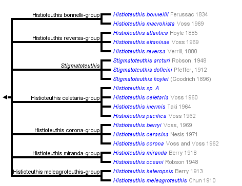


This tree diagram shows the relationships between several groups of organisms.
The root of the current tree connects the organisms featured in this tree to their containing group and the rest of the Tree of Life. The basal branching point in the tree represents the ancestor of the other groups in the tree. This ancestor diversified over time into several descendent subgroups, which are represented as internal nodes and terminal taxa to the right.
You can click on the root to travel down the Tree of Life all the way to the root of all Life, and you can click on the names of descendent subgroups to travel up the Tree of Life all the way to individual species.
For more information on ToL tree formatting, please see Interpreting the Tree or Classification. To learn more about phylogenetic trees, please visit our Phylogenetic Biology pages.
close boxIntroduction
The Histioteuthidae contains mostly weakly muscled species of moderate size (up to 33 cm ML). In general they have very long, thick arms and short mantles with small rounded fins. One of the most distinctive features of these squid is the different size and orientation of the eyes. The left eye is much larger than the right eye and has a semitubular, rather than a hemispherical, shape. This larger eye, while mobile, generally is directed posterodorsally as indicated by the distribution of iridophores on its outer surface. In this position the eye, on an obliquely oriented squid, is directed vertically upward. During the day in middepths this eye, presumably looks for silhouettes of animals against the dim downwelling light. The right eye has a normal shape and points laterally and slightly downward. Learn more about the eyes here.

Figure. Left and right side views of Stigmatoteuthis hoylei as the squid holds onto large forceps in a ship-board aquarium.
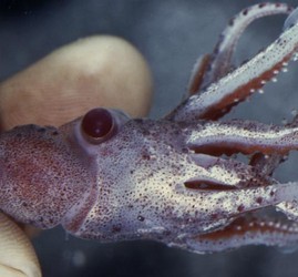
Figure. Posterodorsal view of S. hoylei held by hand. The view is directly into the upward-looking large eye but the smaller eye cannot be seen.
The systematics of the Histioteuthidae is well known compared to most families of oceanic squids due to the excellent and detailed work of N. Voss and her colleagues. Most of the information presented here on the species of this family comes directly (often verbatum) from their works. For a fuller account of this family the viewer should consult these papers, especially Voss, 1969 and Voss, Nesis and Rodhouse, 1998.
Brief diagnosis:
A member of the histioteuthid families ...
- with large, characteristic, integumental photophores on head, arms and mantle.
Characteristics
- Arms
- Inner web between oral surfaces of arms weakly to strongly developed. Occasionally outer web between aboral surfaces of arms also present.
- Inner web between oral surfaces of arms weakly to strongly developed. Occasionally outer web between aboral surfaces of arms also present.
- Head
- Left eye much larger than right eye.
- Left eye much larger than right eye.
- Photophores
- Ventral surfaces of mantle, head and arms (but not funnel) with anteriorly directed, compound photophores with red color filters and unique morphology. Click on an image to view larger version & data in a new window
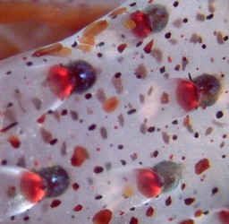
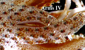
Figure. Ventral view of photophores on arm IV, S. hoylei, off Hawaii. Photograph by R. Young.
The histological structure of a photophore can be seen here.
- Ventral surfaces of mantle, head and arms (but not funnel) with anteriorly directed, compound photophores with red color filters and unique morphology.
- Gladius
- Posterior end of gladius with a cupped coil.
- Posterior end of gladius with a cupped coil.
- Viscera
- Primary and secondary efferent blood vessels of the gills highly pigmented. This feature appears to be characteristic of histioteuthids and is found rarely in other oegopsids (e.g. Ancistrocheirus, Architeuthis). Click on an image to view larger version & data in a new window
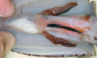
Figure. Ventral view of the opened mantle cavity of Histioteuthis reversa, mature male, showing the pigmented blood vessels of the gills. Photograph by M. Vecchione.
- Primary and secondary efferent blood vessels of the gills highly pigmented. This feature appears to be characteristic of histioteuthids and is found rarely in other oegopsids (e.g. Ancistrocheirus, Architeuthis).
Comments
The pattern of photophores is important in identifying species and species groups. Some of the basic patterns and the terminology associated with them can be found here.
Comparisons among species groups
Voss (1969) and Voss, et al (1998) have recognized subspecies in several Histioteuthis species. Since designation of either species or subspecies in this family is based on arbitrary criteria concering the degree of difference between the taxa, we prefer to consider their subspecies as species. The present 19 taxa of the genus are placed into 7 species groups on the basis of similarity (Voss, et al., 1998). The table below examines the distribution of 9 important systematic characteristics that separate the species groups, and in some cases, species within a group.
| Group | Species | Photop. series, arms IV | Photop. sizes, ant. mantle | Photop., arm-tip, simple | Photop., arm-tip, with gap | Photop. head pattern | Photop. no. in Basal Row | Tubercles | Inner web | Paired penes |
|---|---|---|---|---|---|---|---|---|---|---|
| reversa | H. atlantica | 4 | intermixed | present | absent | ? | ? | absent | shallow | absent |
| H. eltaninae | 3 | intermixed | absent | absent | ? | ? | absent | shallow | absent | |
| H. reversa | 4 | intermixed | absent | absent | ? | ? | absent | shallow | absent | |
| Stigmatoteuthis | S. arcturi | 3 | uniform | absent | absent | Type 1a | 8 | absent | shallow | present |
| S.dofleini | 3 | uniform | absent | absent | Type 1a | 8 | absent | shallow | present | |
| S.hoylei | 3 | uniform | absent | absent | Type 1a | 8 | absent | shallow | present | |
| bonnellii | H. bonnellii | 3 | uniform | present | absent | Type 1b | 7 | absent | deep | absent |
| H. macrohista | 3 | uniform | present | absent | ? | ? | absent | deep | absent | |
| celetaria | H. celetaria | 3 | uniform | absent | present | Type 1b | 9 | absent | shallow | absent |
| H. inermis | 3 | uniform | absent | absent | Type 1b | 10 | absent | shallow | absent | |
| H. pacifica | 3 | uniform | absent | present | Type 1b | 9 | absent | shallow | absent | |
| H. sp. A | 3 | uniform | absent | present | Type 1b | 9 | absent | shallow | absent? | |
| corona | H. berryi | 4 | uniform | absent | absent | Type 2b | 7 | absent | shallow | absent |
| H. cerasina | 3 | uniform | absent | absent | Type 2a | 7 | absent | shallow | absent | |
| H. corona | 3 | uniform | absent | absent | Type 2a | 7 | absent | shallow | absent | |
| miranda | H. miranda | 5 | uniform | absent | absent | ? | ? | present | shallow | absent |
| H. oceani | 6 | uniform | absent | absent | ? | ? | present | shallow | absent | |
| meleagroteuthis | H. meleagroteuthis | 8-9 | uniform | absent | absent | ? | ? | present | shallow | absent |
| H. heteropsis | 8-10 | uniform | absent | absent | ? | ? | absent | shallow | absent |
Some of the characters in the table above are illustrated here:
Figures. Left to right:
- Photop. series, arms IV - Ventral view of arms IV showing photophores in three series, Stigmatoteuthis hoylei, off Hawaii. Photograph by R. Young.
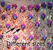
- Photop., sizes, ant. mantle - Ventral view of mantle, Histioteuthis reversa , showing mantle photophores of two different sizes. Photograph by R. Young.
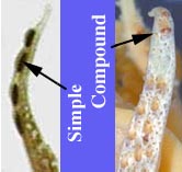
- Photop., arm-tip, simple - Lateral view of arm tip, H. atlantica, showing simple (dark), elongate photophores of the aboral surface. Photograph by E. McSweeny.
- photop., arm-tip, with gap - Ventral view of arm IV, H. sp. A, showing group of arm-tip photophores separated from proximal photophores by a gap. Photograph by R. Young.
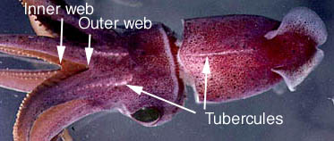
- Inner web; tubercules - Dorsal view of H. oceani, off Hawaii showing web between arms and rows of tubercules. Photograph by R. Young.
- Photop., head pattern; Photop., no. in Basal Row - See "Comments" above.
- Paired penes - All of these structures are paired: penis, needhams sac, spermatophore glands and ducts.
Nomenclature
A list of all nominal genera and species in the Histioteuthidae can be found here. The list includes the current status and type species of all genera, and the the current status, type repository and type locality of all species and all pertinent references.
Life History
Paralarvae have not been positively identified to species. Paralarvae, however, are easily recognized as being histioteuthids by the long arms, in combination with the very large arm suckers relative to the very small tentacular club suckers. The abrupt transition from the paralarval to the juvenile stage in histioteuthids involves pronounced morphological changes. This makes paralarval identification difficult unless intermediates in the changeover period can be found.
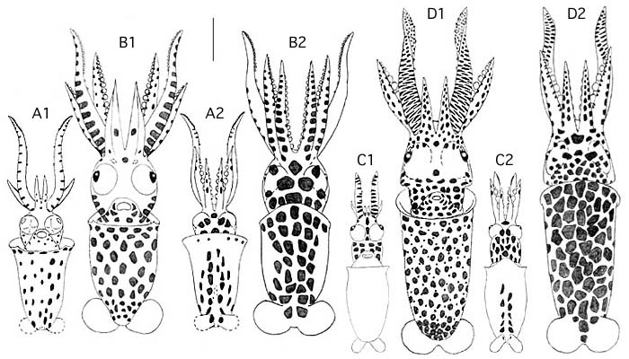
Figure. Ventral and dorsal views of paralarvae of two sizes of two species of histioteuthids. A, B - Probably growth stages of Stigmatoteuthis. hoylei, based on relative abundance. A1, A2 - 2.0 mm ML, B1, B2 - 2.9 mm ML. C, D - Probably growth stages of either H. oceani or H. cerasina, the other two common Hawaiian histioteuthids. C1, C2 - 1.8 mm ML, D1, D2 - 3.6 mm ML. All were taken off Hawaii. Original drawings by R. young. The bar is 1 mm.
At or near sexual maturity many species of histioteuthids exhibit morphological change often involving the addition of photophores and in at least one case (H. reversa) a change in body proportions.
Behavior
Histioteuthids observed from submersibles characteristically have the mantle oblique with all arms curled above the head which, in long-armed species, can give the impression that the squid is tied in a knot (see photograph below). Presumably this posture is in response to disturbance by the submersible. In addition to the usual function of fins, the small fins at the posterior tip of the mantle form a circle and swimming waves from one will continue onto the other causing the squid to rotate about its axis (Hunt, 1996).
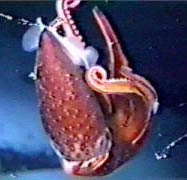
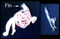
Figure. Left - In situ video frames of a histioteuthid (probably H. reversa), depth unknown. Right - Two video frames of Stigmatoteuthis arcturi, 800 m, Bahamas, show a hovering S. arcturi with "knotted arms" on the left, then the same squid slowly swimming away on the right. Pictures provided by M. Vecchione.
References
Hunt, J. C. 1996. The behavior and ecology of midwater cephalopods from Monterey Bay: Submersible and laboratory observations. Ph. D. Dissertation, Univ. Calif. Los Angeles. 231 pp.
Voss, N. A. 1969. A monograph of the Cephalopoda of the North Atlantic: The family Histioteuthidae. Bull. Mar. Sci., 19: 713-867.
Voss, N.A., K. N. Nesis, P. G. Rodhouse. 1998. The cephalopod family Histioteuthidae (Oegopsida): Systematics, biology, and biogeography. Smithson. Contr. Zool., 586(2): 293-372.
Voss, N. A., S. J. Stephen and Zh. Dong. 1992. Family Histioteuthidae. Smithson. Contr. Zool., No. 513: 73-91.
Title Illustrations

| Scientific Name | Stigmatoteuthis hoylei |
|---|---|
| Location | off Hawaii |
| View | ventrolateral |
| Image Use |
 This media file is licensed under the Creative Commons Attribution-NonCommercial License - Version 3.0. This media file is licensed under the Creative Commons Attribution-NonCommercial License - Version 3.0.
|
| Copyright |
© 1996

|
About This Page
University of Hawaii, Honolulu, HI, USA
National Museum of Natural History, Washington, D. C. , USA
Page copyright © 2016 and
 Page: Tree of Life
Histioteuthidae .
Authored by
Richard E. Young and Michael Vecchione.
The TEXT of this page is licensed under the
Creative Commons Attribution-NonCommercial License - Version 3.0. Note that images and other media
featured on this page are each governed by their own license, and they may or may not be available
for reuse. Click on an image or a media link to access the media data window, which provides the
relevant licensing information. For the general terms and conditions of ToL material reuse and
redistribution, please see the Tree of Life Copyright
Policies.
Page: Tree of Life
Histioteuthidae .
Authored by
Richard E. Young and Michael Vecchione.
The TEXT of this page is licensed under the
Creative Commons Attribution-NonCommercial License - Version 3.0. Note that images and other media
featured on this page are each governed by their own license, and they may or may not be available
for reuse. Click on an image or a media link to access the media data window, which provides the
relevant licensing information. For the general terms and conditions of ToL material reuse and
redistribution, please see the Tree of Life Copyright
Policies.
- Content changed 03 November 2013
Citing this page:
Young, Richard E. and Michael Vecchione. 2013. Histioteuthidae . Version 03 November 2013 (under construction). http://tolweb.org/Histioteuthidae/19782/2013.11.03 in The Tree of Life Web Project, http://tolweb.org/

