


Mastigoteuthis cordiformis
Richard E. Young and Michael Vecchione

Introduction
The original description of M. cordiformis is based on a single male squid, 80 mm ML with intact tentacles, taken in the Indian Ocean near Sumatra. It has been subsequently described from Japan (Sasaki, 1929) and the Philippines (Voss, (1963). This is the largest of the mastigoteuthids reaching a size of 100 cm ML (A. Salcedo-Vargas, personnal communication).
Brief diagnosis:
A mastigoteuthid ...
- without photophores.
- with club bearing very large suckers (ca. 0.5 mm) at proximal end.
- with skin tubercules.
Characteristics
- Arms
- Arms III much longer than arms I.
- Tentacles
- Proximal club suckers much larger than other club suckers.
- Largest proximal suckers 0.5 mm or larger.
- Head
- Beaks. Description of the beaks can be found here.
- Funnel pocket absent.
- Beaks. Description of the beaks can be found here.
- Funnel
- Funnel locking-apparatus ear-shaped with tragus and antitragus.
- Fins
- Fin length (anterior extent to insertion on tail) 75% of ML (Chun, 1910).
- Tubercules
- Mantle and other skin covered with small, conical tubercules arising from round plaques.
- Photophores and pigmentation
- Photophores absent.
- Most pigment in densely packed chromatophores.
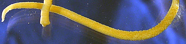
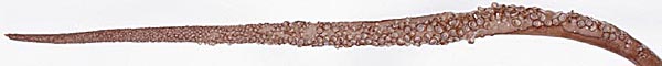
Figure. Oral view of the tentacular club of M. cordiformis. Note the large suckers at the proximal end. Top - Holotype, 80 mm ML. Photograph by R. Young. Bottom - Large specimen (>30 cm ML), fresh, Australian waters. Photograph by Mark Norman.
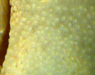
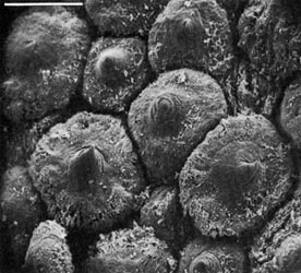
Figure. Left - Ventral view of a portion of the eyelid of M. cordiformis, holotype, 80 mm ML, showing tile-like arrangement of tubercules. Photograph by R. Young. Right - Scanning electron micrograph with an external view of mantle tubercles of M. cordiformis, 87 mm ML, Philippine waters, 14°N, 121°E. The tubercules are very similar to those of M. hjorti. Photograph from Roper and Lu (1990).
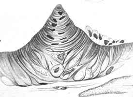
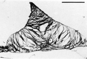
Figure. Left - Histological section through a skin tubercule of M. cordiformis, holotype. Most of the epithelium has been lost except along one flank of the tubercule. Drawing from Chun (1910). Right - Cross-section through a tubercule of M. cordiformis using scanning electron microscopy. Photograph from Roper and Lu (1990).
Comments
More details of the description can be found here.
The descriptions of Chun, Sasaki and Voss contain differences in the shape of the funnel locking-apparatus and the structure of the suckers. The significance of these differences remains to be determined. M. latipinna Sasaki, 1916 (see description under "Species of doubtful validity..." in the Nomenclature section of the family page) is probably a junior synonym of M. cordiformis.
M. cordiformis is most similar to M. hjorti but differs in the absence of ocular photophores and the presence of enlarged proximal club suckers among other features.
Distribution
Type locality: Indian Ocean south of Sumatra at 0°15'N, 98°8'E. Also known from southern Japanese (Sasaki, 1929) and Philippine (Voss, 1963) waters.
References
Chun, C. 1910. Die Cephalopoden. Oegopsida. Wissenschaftliche Ergebnisse der Deutschen Tiefsee Expedition auf dem Dampfer "Valdivia" 1898-1899, 18(1):1-401.
Roper, C.F.E. and C.C. Lu 1990. Comparative morphology and function of dermal structures in oceanic squids (Cephalopoda). Smithson. Contr. Zool., No. 493: 1-40.
Sasaki, M. 1929. A Monograph of the Dibranchiate Cephalopods of the Japanese and Adjacent Waters. Journal of the College of Agriculture, Hokkaido Imperial University, 20(supplement):357 pages.
Voss, G. L. 1963. Cephalopoda of the Philippine Islands. Bull. U. S. Nat. Mus., 234: 1-180.
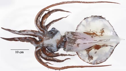
Title Illustrations

| Scientific Name | Mastigoteuthis cordiformis |
|---|---|
| Location | South Pacific |
| View | Ventral |
| Size | 325 mm ML |
| Copyright | © 2004 Mark Norman |
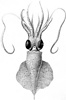
| Scientific Name | Mastigoteuthis cordiformis |
|---|---|
| Reference | Chun, C. 1910. Die Cephalopoden. Oegopsida. Wissenschaftliche Ergebnisse der Deutschen Tiefsee Expedition auf dem Dampfer "Valdivia" 1898-1899, 18(1):1-401. |
| View | Ventral |
| Size | 83 mm ML (including tail). |
| Type | Holotype |
About This Page
Richard E. Young

University of Hawaii, Honolulu, HI, USA
National Museum of Natural History, Washington, D. C. , USA
Page copyright © 2004 Richard E. Young and
All Rights Reserved.
- First online 16 July 2004
- Content changed 19 November 2007
Citing this page:
Young, Richard E. and Vecchione, Michael. 2007. Mastigoteuthis cordiformis . Version 19 November 2007. http://tolweb.org/Mastigoteuthis_cordiformis/19510/2007.11.19 in The Tree of Life Web Project, http://tolweb.org/


