


Mastigoteuthis microlucens
Richard E. Young, Michael Vecchione, and Annie Lindgren
Introduction
Mastigoteuthis microlucens, previously listed here as Mastigoteuthis sp. A, is the most common species of the genus around the main Hawaiian Islands. It has numerous tiny photophores that lie beneath the outer layer of integumental chromatophores. The photophores are so small that they cannot be recognized as photophores without the aid of a microscope.
Brief diagnosis:
A member of the M. magna group with ...
- minute integumental photophores.
- a tropical Pacific habitat.
Characteristics
- Arms
- Arms II moderate in length, 46% of ML, 69% of arms II length.
- Large arm suckers generally with closely-set, rounded teeth on the distal margin, occasionallly fused; sometimes smaller, more proximal teeth with narrower, more distinctly separated teeth. Click on an image to view larger version & data in a new window
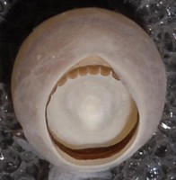
Figure. Oral view of an arm sucker (sucker 26, arm III) of M. microlucens, paratype. Photograph by R. Young.
Scanning electron micrographs of the arm and tentacle suckers can be seen here.
- Head
- Funnel
- Funnel component of the funnel/mantle locking-apparatus flask-shaped with narrow stem.
- Mantle component without a nostril. Click on an image to view larger version & data in a new window
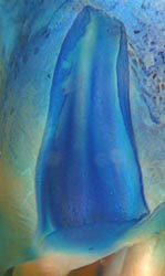
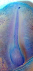
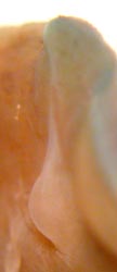
Figure. Funnel-mantle locking-apparatus of M. microlucens, holotype, Equatorial Pacific. Left - Frontal view of funnel component. Middle - Frontal view of mantle component. Right - Side view of mantle component. Blue stain = methylene blue. Photographs by R. Young.
- Photophores
- Minute ("lens" diameter less than 0.1 mm) integumental photophores present but unrecognizable to naked eye due to small size and structure.
- Integumental photophores present on aboral surfaces of all arms, on dorsal and ventral surfaces of Head, mantle and fins, on ventral surface of funnel.
- Integumental photophores lie beneath the surface layer of chromatophores.
- Eyelid photophores absent. Click on an image to view larger version & data in a new window
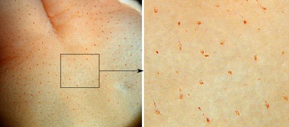
Figure. Ventral view of the head of M. microlucens, holotype, Equatorial Pacific. Outer chromatophores were lost during capture leaving the scattered photophores visible. Enlargement at right allows photophores to be recognized by the presence of small, oval lenses. Photographs by R. Young.
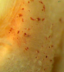
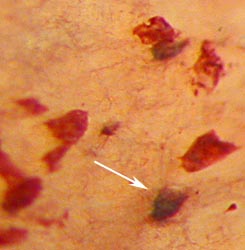
Figure. Photophores of M. microlucens. Left - Anteriolateral view of the head near the eye opening looking at the lenses of the photophores. Right - High magnification of two photophores (one indicated by the arrow) and a few, lighter-red chromatophores that are more superficially located from a damaged region of the side of the head, 160 mm ML, Hawaiian waters.
Comments
More details of the description can be seen here.
We identify structures here as photophores on the basis of a dark, pigmented "cup" and a spherical, whitish "lens" and their distribution as a separate layer beneath most chromatophores. No bioluminescence has been observed or attempted to be observed. M. microlucens is unique among members of the M. magna-group in having photophores. If the surface chromatophore layer is intact, the deeper lying photophores can be difficult to recognize even under a microscope. Mastigoteuthis sp. A also differs from M. magna in the more elongate shape of the posterior bulb of the funnel locking-apparatus, dentition of the arm suckers and details of the beaks.
Molecular Characteristics
M. microlucens is most closely related to M. magna based on morphological evidence (see the M. magna-group page). Molecular data indicate substantial divergence across the three genes examined (see Table below) and confirms that M. microlucens is a separate species from M. magna. Hebert et al (2003) hypothesized a 2% rate of divergence in the COI locus as a general guideline for identifying distinct species, here the rate of divergence between M. microlucens and the morphologically similar M. magna was almost 10%. The more morphologically distinct M. hjorti had a COI sequence divergence rate 12% with M. microlucens, only slightly greater than that of M. magna which further emphasizes the surprising result of 10% difference seen with M. magna (Young, Lindgren and Vecchione, submitted) (see also the Discussion of Phylogenetic Relationships on the Mastigoteuthis page).
| Locus | Base pairs | M. magna | M. hjorti | |
| M. microlucens | COI rRNA | 658 | 9.89% | 11.7% |
| M. microlucens | 16S rRNA | 528 | 3.40% | 6.02% |
| M. microlucens | 12S rRNA | 404 | 5.13% | 7% |
More details on the squid that provided the molecular data can be found here.
Life History
At 5 mm ML M. microlucens paralarvae are separated from similar-sized paralarvae of M. famelica, the only other common mastigoteuthid in Hawaiian waters, by the broader shape, larger eyes, more posterior digestive gland and tentacular suckers and chromatophores restricted to the distal end of the tentacle. At 7 mm ML the presence of anterior fin lobes also distinguishes M. microlucens. At larger sizes, the slender shape, elongate fins and, by at least 17 mm ML, skin tubercules easily distinguish M. famelica. The differences between the two species diminish at sizes below 4.5 mm ML. The position of the digestive gland is useful to at least 3.5 mm ML. The smallest M. microlucens paralarva captured had a mantle length of 2.5 mm ML.
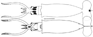
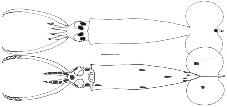
Figure. Ventral and dorsal views of M. microlucens paralarvae, Hawaiian waters. Top - 4.6 mm ML. Bottom - 7.2 mm ML. Scale bars = 1 mm. Drawings from Young (1991) labeled as M. inermis.
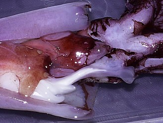
Figure. Mature male of M. microlucens, 215 mm ML, Hawaiian waters, showing large penis containing white spermatophores and two whitish sacs of the distal reproductive tract emerging from the genital sac. Photograph by R. Young.
Distribution
Geographical Distribution
Type locality: Equatorial Pacific south of Hawaii.
We have captured M. microlucens in the region of the Hawaiian Archipelago to about ca. 26°N and south to the equator. This is the most common mastigoteuthid encountered in the region of the high (main) Hawaiian Islands.
Vertical Distribution
The vertical distribution of M. microlucens in Hawaiian waters was examined by Young (1978). During the day most captures came from depths of 675-870 m; nighttime captures came from 255 to 725 m with most squid taken between 250 and 450 m.
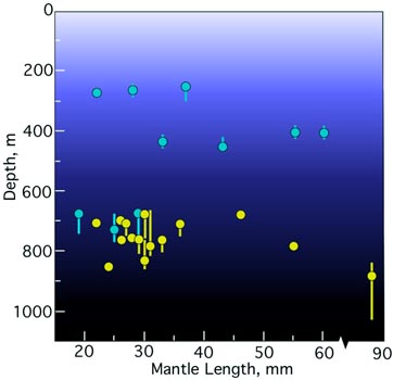
Figure. Vertical distribution chart of M. microlucens. Captures were made with both open and opening/closing trawls. Bar - fishing depth-range of opening/closing trawl. Circle - Modal fishing depth for either trawl. Blue-filled circle - Night capture. Yellow-filled circle - Day capture. Note the breaks in the x-axis. Chart modified from Young (1978, labeled as M. inermis).
References
Hebert, P.D., S. Ratnasingham, and J.R. de Waard. 2003. Barcoding animal life: cytochrome c oxidase subunit I divergences among closely related species. Proc. R. Soc. Lond. B., 270: S96-S99
Young, R. E. 1978. Vertical distribution and photosensitive vesicles of pelagic cephalopods from Hawaiian waters. Fish. Bull., 76: 583-615.
Young, R. E. (1991). Chiroteuthid and related paralarvae from Hawaiian waters. Bull. Mar. Sci., 49: 162-185.
Young, R. E., A. Lindgren and M. Vecchione. 2008. Mastigoteuthis microlucens, a new species of the squid family Mastigoteuthidae (Mollusca: Cephalopoda). Proc. Biol. Soc. Wash. 121(2): 276-282.
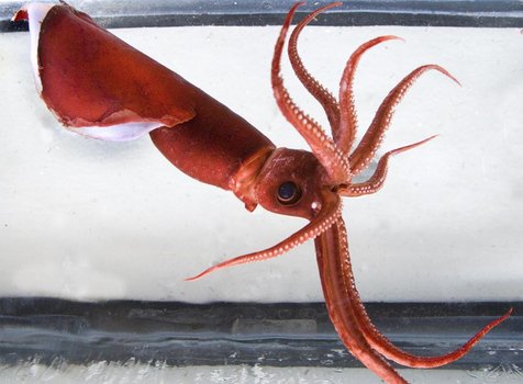
Title Illustrations

| Scientific Name | Mastigoteuthis microlucens |
|---|---|
| Location | Off Keahole Point, Hawaii Island |
| Reference | Modified from: Young, R. E., A. Lindgren and M. Vecchione. 2008. Mastigoteuthis microlucens, a new species of the squid family Mastigoteuthidae (Mollusca: Cephalopoda). Proc. Biol. Soc. Wash. 121(2): 276-282. |
| Creator | Michael Darden, photographer |
| Specimen Condition | Live Specimen |
| Sex | Female |
| Life Cycle Stage | Immature |
| View | Side |
| Size | 135 mm ML |
| Copyright | © West Hawaii Today |
About This Page
Richard E. Young

University of Hawaii, Honolulu, HI, USA
National Museum of Natural History, Washington, D. C. , USA
Ohio State University, Columbus, Ohio, USA
Page copyright © 2007 Richard E. Young, , and
 Page: Tree of Life
Mastigoteuthis microlucens .
Authored by
Richard E. Young, Michael Vecchione, and Annie Lindgren.
The TEXT of this page is licensed under the
Creative Commons Attribution-NonCommercial License - Version 3.0. Note that images and other media
featured on this page are each governed by their own license, and they may or may not be available
for reuse. Click on an image or a media link to access the media data window, which provides the
relevant licensing information. For the general terms and conditions of ToL material reuse and
redistribution, please see the Tree of Life Copyright
Policies.
Page: Tree of Life
Mastigoteuthis microlucens .
Authored by
Richard E. Young, Michael Vecchione, and Annie Lindgren.
The TEXT of this page is licensed under the
Creative Commons Attribution-NonCommercial License - Version 3.0. Note that images and other media
featured on this page are each governed by their own license, and they may or may not be available
for reuse. Click on an image or a media link to access the media data window, which provides the
relevant licensing information. For the general terms and conditions of ToL material reuse and
redistribution, please see the Tree of Life Copyright
Policies.
- First online 19 November 2007
- Content changed 06 August 2008
Citing this page:
Young, Richard E., Vecchione, Michael, and Lindgren, Annie. 2008. Mastigoteuthis microlucens . Version 06 August 2008 (under construction). http://tolweb.org/Mastigoteuthis_microlucens/65304/2008.08.06 in The Tree of Life Web Project, http://tolweb.org/


