


Myopsida
Michael Vecchione and Richard E. Young


This tree diagram shows the relationships between several groups of organisms.
The root of the current tree connects the organisms featured in this tree to their containing group and the rest of the Tree of Life. The basal branching point in the tree represents the ancestor of the other groups in the tree. This ancestor diversified over time into several descendent subgroups, which are represented as internal nodes and terminal taxa to the right.
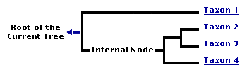
You can click on the root to travel down the Tree of Life all the way to the root of all Life, and you can click on the names of descendent subgroups to travel up the Tree of Life all the way to individual species.
For more information on ToL tree formatting, please see Interpreting the Tree or Classification. To learn more about phylogenetic trees, please visit our Phylogenetic Biology pages.
close boxIntroduction
Myopsid squids are neritic, often in very shallow water, or upper slope demersal species. Many species are strong swimmers, occur in large schools and are fished commercially for food. The Loliginidae contains many species some of which reach a rather large size (at least 90 cm ML in Loligo forbesii) but those in Pickfordiateuthis, are dwarf species where males may mature at less than 14 mm ML (Brachoniecki, 1996). The Australiteuthidae contains a single species that is also a dwarf with males that mature as small as 17 mm ML (Lu, 2005).
Brief diagnosis:
A decapodiform ...
- with corneal membranes covering eye lenses.
- without secondary eyelids.
- with well-developed gladius.
Characteristics
- Arms
- Suckers of arms (and tentacles) with circularis muscles (unknown in Australiteuthidae).
- Tentacles
- Club without proximal (= carpal) locking-apparatus. More details of the typical club can be found here.
- Head
- Head with tentacle pocket.
- Eyes with corneal membranes covering lenses.
- Eyes without secondary (= ventral) eyelid.
- Funnel
- Funnel without lateral adductor muscles.
- Mantle
- Mantle locking-apparatus extends to mantle edge (except Australiteuthidae).
- Shell
- Shell a gladius, extending the full length of the mantle.
- Viscera
- Gills with branchial canal (except Pickfordiateuthis, unknown in Australiteuthis).
- Right oviduct absent.
- Females with accessory nidamental glands.
- Eggs
- Eggs, where known, attached to substrate.
- Embryo with large external yolk sac.
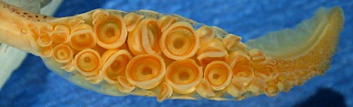
Figure. Oral view of the tentacular club of Loligo plei, 105 mm ML, preserved. Photograph by R. Young.
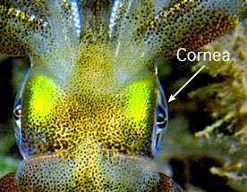
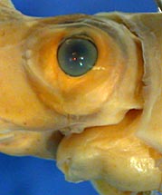
Figure. Left - Dorsal view of the head of Sepioteuthis sp. (Loliginidae) showing cornea covering lens. Photograph by Mark Norman. Right - Ventrolateral view of the head of Lollicuncula diomedeae showing the absence of a ventral eyelid. Photograph by R. Young.

Figure. Diagramatic cross-section through gills. Drawing modified from Naef (1921-23).
Discussion of Phylogenetic Relationships
The two families of the Myopsida appear to be closely related. The Australiteuthidae differs from the Loliginidae primarily in the structure of the funnel/mantle locking-apparatus and the position of the mantle component which does not reach the mantle margin. A number of features of phylogenetic importance in the Australiteuthidae, however, are not known: the presence or absence of a branchial canal, an anterior eye pocket, circularis muscles in the suckers, an interstellate connective; the location of spermathecae; the symmetry of the gills; the type and place (pelagic or benthic) of deposition of egg masses and the position of the intestine relative to the cephalic vein and the vena cavae.
References
Brakoniecki, T. F. 1996. A revision of the genus Pickfordiateuthis Voss, 1953 (Cephalopoda; Myopsida). Bull. Mar. Sci., 58: 9-28.
Lu, C. C. 2005. A new family of myopsid squid from Australasian waters (Cepahlopoda: Teuthida). P. 71-82. In: Chotiyaputta, C., E. M. C. Hatfield and C. C. Lu (editors). Cephalopod biology, recruitment and culture. International Cephalopod Symposium and Workshop, 17-21 Feb. 2003. Research Bulletin, Phyuket Marine Biological Center, No. 66, Published by the Center Phuket, Thailand, July 2005, 365 pp.
Naef, A. 1921-1923. Die Cephalopoden. Fauna e Flora del Golfo di Napoli, Monographie 35, Vol I, Parts I and II, Systematik, pp 1-863.
Title Illustrations

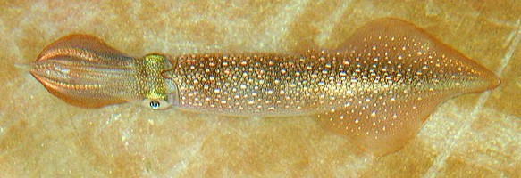
| Scientific Name | Doryteuthis pealeii |
|---|---|
| Specimen Condition | Live Specimen |
| View | Dorsal |
| Size | 105 mm ML |
| Image Use |
 This media file is licensed under the Creative Commons Attribution-NonCommercial License - Version 3.0. This media file is licensed under the Creative Commons Attribution-NonCommercial License - Version 3.0.
|
| Copyright |
©

|
About This Page
National Museum of Natural History, Washington, D. C. , USA
University of Hawaii, Honolulu, HI, USA
Page copyright © 2019 and
 Page: Tree of Life
Myopsida .
Authored by
Michael Vecchione and Richard E. Young.
The TEXT of this page is licensed under the
Creative Commons Attribution-NonCommercial License - Version 3.0. Note that images and other media
featured on this page are each governed by their own license, and they may or may not be available
for reuse. Click on an image or a media link to access the media data window, which provides the
relevant licensing information. For the general terms and conditions of ToL material reuse and
redistribution, please see the Tree of Life Copyright
Policies.
Page: Tree of Life
Myopsida .
Authored by
Michael Vecchione and Richard E. Young.
The TEXT of this page is licensed under the
Creative Commons Attribution-NonCommercial License - Version 3.0. Note that images and other media
featured on this page are each governed by their own license, and they may or may not be available
for reuse. Click on an image or a media link to access the media data window, which provides the
relevant licensing information. For the general terms and conditions of ToL material reuse and
redistribution, please see the Tree of Life Copyright
Policies.
- First online 20 September 2005
- Content changed 29 August 2016
Citing this page:
Vecchione, Michael and Richard E. Young. 2016. Myopsida . Version 29 August 2016. http://tolweb.org/Myopsida/52670/2016.08.29 in The Tree of Life Web Project, http://tolweb.org/

