The mantle of L. grimaldii is covered by a dense layer of dermal cushions. The layer ends abruptly (see arrow in photograph below) in the anterior half of the fins presumably at about the point where the muscular mantle ends.

Click on an image to view larger version & data in a new window

Figure. Dermal cushions of mantle of L. grimaldii, 617 mm ML, fresh. Photograph by D. Stevens.
Click on an image to view larger version & data in a new window
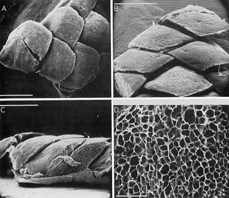
Figure. Scanning electron micrographs of the cushions of L. grimaldii and their structure, scale bar in A-C = 1 mm, in D = 0.1 mm. A - Dermal cushions in normal position on mantle with anterior cushions overlapping posterior ones. B - Cushions viewed posteriorly. C - Longitudinal section through one scale and part of another. Note the spongy appearance. D - Closer view of the spongy structure of the cushion showing numerous vacuoles. Photographs from Roper and Lu (1990).
About This Page

University of Hawaii, Honolulu, HI, USA
National Museum of Natural History, Washington, D. C. , USA
Page copyright © 1999 and
 Page: Tree of Life
Lepidoteuthis grimaldii: Dermal Cushions
Authored by
Richard E. Young and Michael Vecchione.
The TEXT of this page is licensed under the
Creative Commons Attribution-NonCommercial License - Version 3.0. Note that images and other media
featured on this page are each governed by their own license, and they may or may not be available
for reuse. Click on an image or a media link to access the media data window, which provides the
relevant licensing information. For the general terms and conditions of ToL material reuse and
redistribution, please see the Tree of Life Copyright
Policies.
Page: Tree of Life
Lepidoteuthis grimaldii: Dermal Cushions
Authored by
Richard E. Young and Michael Vecchione.
The TEXT of this page is licensed under the
Creative Commons Attribution-NonCommercial License - Version 3.0. Note that images and other media
featured on this page are each governed by their own license, and they may or may not be available
for reuse. Click on an image or a media link to access the media data window, which provides the
relevant licensing information. For the general terms and conditions of ToL material reuse and
redistribution, please see the Tree of Life Copyright
Policies.


