Larvae of some species have ciliated patches, others do not. Those of Multicotyle purvisi have a circle of four patches at the anterior margin of the posterior sucker and a circle of six at the posterior end, those of Cotylogaster occidentalis have an anterior ring of eight and a posterior ring of six. Larvae of Aspidogaster conchicola, Lobatostoma manteri, Rugogaster hydrolagi and Multicalyx elegans lack cilia. Larvae of some species hatch from the eggs, others do not; I discuss one of each in greater detail (Rohde, 1972, 1994, other references therein).
Multicotyle purvisi
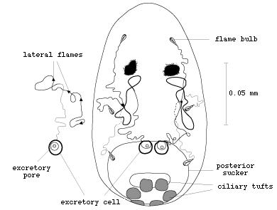
Multicotyle purvisi is a species that hatches before entry into the host. It infects freshwater snails and turtles in Southeast Asia. It has a pharynx and short caecum, one pair of eyes, a posterior sucker, a circle of six ciliated cells at the posterior end, and four ciliated cells at the anterior margin of the posterior sucker (Fig. 1).


Figure 1. Larva of Multicotyle purvisi. Note four anterior and six posterior ciliary tufts (redrawn from Rohde, 1972).
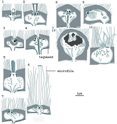
The nucleus of each ciliated cell is located in a pocket of cytoplasm covered by tegument. The surface tegument is drawn out into many thin processes, the so-called microfila (Fig.2).


Figure 2. Section through ciliated cell of larval Multicotyle purvisi. Note nucleus in cytoplasmic pocket located below the tegument, cilia with single oblique rootlets, and microfila (thin surface outgrowths of the tegument) (redrawn from Rohde, 1972).
The two excretory bladders are located dorsally, at the anterior margin of the posterior sucker, connected to protonephridial ducts and three flame bulbs on each side of the body; the ducts contain ciliary tufts as well, the so-called lateral flames (Fig.3).


Figure 3. Larva of Multicotyle purvisi with protonephridial (excretory) system (redrawn from Rohde, 1972).
The nervous system is of great complexity, consisting of a great number of longitudinal nerves (connectives) connected by circular commissures. The brain (cerebral commissure) is located dorsally, in the anterior part of the body, the eyes dorsally attached to it. A nerve from the main connective enters the pharynx and also supplies the intestine. Posteriorly, the main connective enters the sucker (Fig.4).


Figure 4. Larva of Multicotyle purvisi with nervous system (redrawn from Rohde, 1972).
Sensory receptors are scattered over the ventral and dorsal surface, the largest numbers occurring on the ventral surface, at the anterior end and on the posterior sucker. Electron-microscopic studies revealed 13 types of receptors (Figs. 5-7) (Rohde and Watson, 1990a, b, c).


Figure 5. Sensory receptors of larval Multicotyle purvisi (redrawn from Rohde and Watson, 1990a,b).
- uniciliate receptors with one electron-dense collar and a cilium about 0.6 um long;
- uniciliate receptors with two electron-dense collars and a cilium containing dense cytoplasm around the ring of peripheral microtubule doublets;
- uniciliate receptors with two electron-dense collars and large reticulate extensions that extend into the receptor;
- uniciliate receptors with two electron-dense collars and a long cross-striated ciliary rootlet;
- uniciliate receptors that contain a cilium with many single microtubules and doublets;
- uniciliate receptors with two electron-dense collars and a cilium at least 6 um long;
- uniciliate receptors with two electron-dense collars and a cilium about 1.3 um long;
- a pair of non-ciliate receptors in the mouth cavity with two to several electron- dense collars, a long cross-striated ciliary rootlet and some fibres that diverge from the basal body;
- four pairs of non-ciliate receptors in the ventral wall of the posterior sucker and in the tegument ventral to the sucker with many electron-dense collars and a large, round-ovoid ciliary rootlet that diverges from the basal body, is cross-striated in its proximal and has many dense structures in its distal part;
- two large non-ciliate bulbous receptors in the lateral surface tegument anterior to the sucker, with some long cytoplasmic processes and much dense material in their apical parts;
- an unpaired receptor with a bulbous projection at the posterior end, with many vesicles and some long cytoplasmic processes
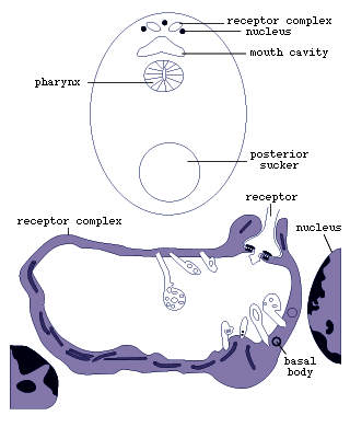
- two anterior receptor complexes dorsal to the mouth cavity, each
consisting of two dendrites, one forming a large cavity with at least
10 short modified cilia, the other penetrating into the cavity of
the former and possessing a single cilium (Fig.6);
 Click on an image to view larger version & data in a new window
Figure 6. Receptor complex of larval Multicotyle purvisi (redrawn from Rohde and Watson, 1990c).
- paired pigmented photoreceptors (ocelli) each consisting
of a pigment cell and two rhabdomeres with perikarya located outside
the ocellus (Fig.7).
 Click on an image to view larger version & data in a new window
Figure 7. Ocellus (eye, photoreceptor) of larval Multicotyle purvisi (redrawn from Rohde and Watson, 1991b).
Lobatostoma manteri
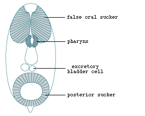
Lobatostoma manteri is a species that does not hatch. The larva lacks cilia and eyes, but resembles the larva of M. purvisi in most other details, possessing a pharynx, short caecum, posterior sucker, and two dorso-posterior excretory bladders. In addition, it has an anterior "false sucker" and a short posterior appendage (Fig. 8) (Rohde, 1973).


Fig. 8. Larva of Lobatostoma manteri (redrawn from Rohde, 1973).
The false sucker is separated from the more posterior tissue by a septum extending to the surface tegument, but is lacks a complete capsule surrounding it, as found in the genuine sucker of the digenean trematodes. Details of the protonephridial and nervous systems are not known. Electron-microscopic studies have shown nine types of receptors, differing in the presence or absence of a cilium, length of the cilium, presence or absence of the ciliary rootlet, and shape of the ciliary rootlet (Fig.9).


Figure 9. Sensory receptors of larval Lobatostoma manteri (redrawn from Rohde and Watson, 1992).
The number of receptor types is smaller than that of Multicotyle purvisi, in particular it lacks eyes and receptor complexes. The difference can be explained by different ways of infecting the host. Whereas Multicotyle purvisi hatches and swims around until inhaled by the snail host, Lobatostoma manteri remains enclosed in the egg shell and is ingested by snails. The larva of Aspidogaster conchicola resembles that of Lobatostoma manteri in many respects, but it has well developed frontal glands which have not been seen in the latter species (Fig.10) (Faust, 1922; Rohde, 1972).


Figure 10. Larva of Aspidogaster conchicola (redrawn after Faust, 1922 from Rohde, 1972).


 Go to quick links
Go to quick search
Go to navigation for this section of the ToL site
Go to detailed links for the ToL site