The early stages of development from the zygote (fertilized egg cell) to the larva has been examined in several species. The egg, as in all parasitic platyhelminths, is ectolecithal, that is, it contains an egg cell and a number of yolk cells. The egg of Multicotyle purvisi is laid at the 1-3 cell stage, whereas the eggs of Aspidogaster conchicola, A. indica and Lobatostoma manteri contain already fully developed larvae (references in Rohde, 1972).
The early embryo of A. conchicola is anteriorly and posteriorly lined by yolk, and the so-called calotte cells at both poles of the egg line the yolk (Fig.1).The yolk is gradually resorbed by the embryo which now fills all of the egg and is lined by the calotte cells. Finally, the false anterior sucker as well as the posterior sucker and posterior appendage appear (Fig.1).

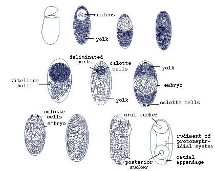
Figure 1. Early development of Aspidogaster conchicola in the egg (redrawn after Voeltzkow, 1888 from Rohde,1972).
Anterior and posterior cells, corresponding to the calotte cells of A. conchicola have also been described for Cotylogaster michaelis. They form an 'embryonic membrane' around the yolk and embryo. In Lobatostoma manteri, as in the other aspidogastreans examined, early cleavage is irregular, that is, no regular pattern in the division of cells can be recognised (Fig.2), and the posterior sucker, which begins to subdivide into alveoli (Fig.3),and the excretory bladder cells appear late in embryogenesis. Interestingly, three to four cells (nuclei with very little cytoplasm) appear in the lumen of the digestive tract of the late embryo. Such cells have also been described in M. purvisi (Fig.4).

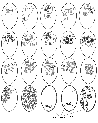
Figure 2. Early development of Lobatostoma manteri in the egg (redrawn from Rohde,1973).

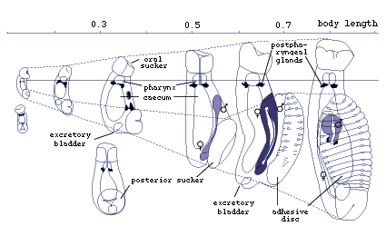
Figure 3. Development of Lobatostoma manteri after hatching. Note the transformation of the posterior sucker into the ventral disc, and - connected with this - the change in the body proportions (redrawn from Rohde,1975).

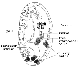
Figure 4. Larva of Multicotyle purvisi just before hatching. Note four free cells in the lumen of the intestine (redrawn from Rohde,1972).
The function of these cells is unknown, and similar cells have not been described from any other flatworm. They are not present in the larvae after hatching.
During further development of M. purvisi, the two bladder cells fuse (Fig.5),

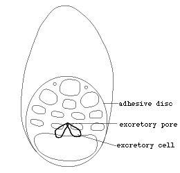
Figure 5. Early juvenile of Multicotyle purvisi, in which the two excretory bladder cells have fused to form a single excretory opening, and in which the posterior sucker has begun to subdivide into alveoles (redrawn from Rohde,1972).
the posterior sucker divides into alveoli, and the male and female reproductive systems develop. The transformation of the larval posterior sucker into the adhesive disc leads to marked changes in the body proportions (Figs.6-8).


Figure 6. Later developmental stages of Multicotyle purvisi, in which the ventral disc is in an advanced stage of development, and in which the male and female reproductive systems are developing (redrawn from Rohde,1972).

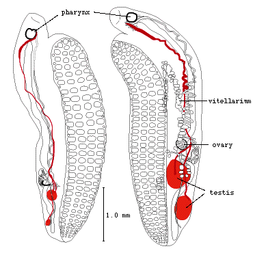
Figure 7. Juveniles of Multicotyle purvisi with well developed ventral discs and reproductive systems (redrawn from Rohde,1972).

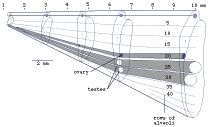
Figure 8. Diagrams of various developmental stages of Multicotyle purvisi to show changes in body proportions (allometric growth) (redrawn from Rohde,1972).
The disc shows at first negative allometric growth (its allometric exponent is less than 1, i.e., it grows relatively more slowly than the whole body) followed by positive allometric growth (allometric component greater than 1, i.e., it grows relatively faster than the whole body). Finally, it shows isometric growth (allometric exponent 1, i.e., it grows as fast as the whole body) (Fig.9).

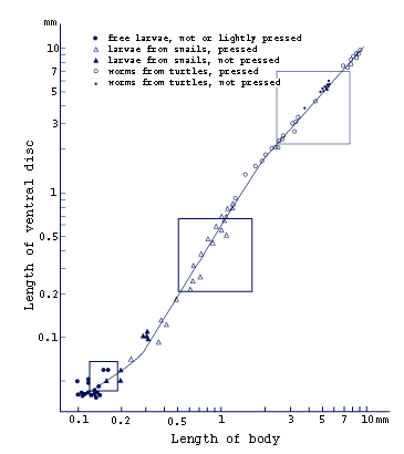
Figure 9. Growth of ventral disc of Multicotyle purvisi. Note the change in the relative length of the ventral disc during growth. The disc shows at first negative allometric growth (allometric exponent less than 1), then positive allometric growth (allometric exponent greater than 1), and finally isometric growth (allometric exponent approximately 1) (redrawn from Rohde,1972).
Similar changes in body proportions have been found in C. manteri and are probably characteristic of all aspidogastreans.


 Go to quick links
Go to quick search
Go to navigation for this section of the ToL site
Go to detailed links for the ToL site