
Click on an image to view larger version & data in a new window
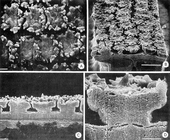
Figure. Scanning electron micrographs of papillate dermal cuchions of Pholidoteuthis massyae 100 mm ML. A - View of the external surface of the tubercules. B - Dermal cuchions viewed posteriorly. Note longitudinally arranged rows. C - Longitudinal section of dermal cuchions showing mushroom-like shape and channels between cushions. D - Enlargement of a section through a cushion. A, C, D scale bars = 0.1 mm. B scale bar = 0.5 mm. Photographs from Roper and Lu (1990).
References
Roper, C.F.E. and C.C. Lu 1990. Comparative morphology and function of dermal structures in oceanic squids (Cephalopoda). Smithson. Contr. Zool., No. 493: 1-40.
About This Page

University of Hawaii, Honolulu, HI, USA
National Museum of Natural History, Washington, D. C. , USA
Page copyright © 1999 and
 Page: Tree of Life
Pholidoteuthis massyae Mantle Dermal Cushions
Authored by
Richard E. Young and Michael Vecchione.
The TEXT of this page is licensed under the
Creative Commons Attribution-NonCommercial License - Version 3.0. Note that images and other media
featured on this page are each governed by their own license, and they may or may not be available
for reuse. Click on an image or a media link to access the media data window, which provides the
relevant licensing information. For the general terms and conditions of ToL material reuse and
redistribution, please see the Tree of Life Copyright
Policies.
Page: Tree of Life
Pholidoteuthis massyae Mantle Dermal Cushions
Authored by
Richard E. Young and Michael Vecchione.
The TEXT of this page is licensed under the
Creative Commons Attribution-NonCommercial License - Version 3.0. Note that images and other media
featured on this page are each governed by their own license, and they may or may not be available
for reuse. Click on an image or a media link to access the media data window, which provides the
relevant licensing information. For the general terms and conditions of ToL material reuse and
redistribution, please see the Tree of Life Copyright
Policies.


