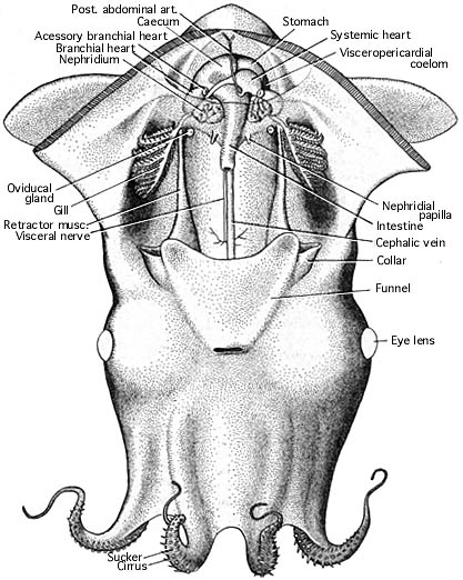
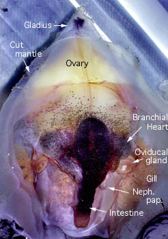
Ventral view of an immature, female V. infernalis with the mantle cavity of cut open. Posteriorly the lining of the mantle cavity has been cut away to better view the visceral nucleus. Note that there is no abdominal septum, no ink sac, no anal flaps. The visceropericardial coelom (dark region surrounding the ventricle, stomach and caecum) is large. Posteriorly, in the ventral midline, the mantle muscle terminates on the conus of the gladius. The photograph on the right shows a mature female. The arrangement of the viscera looks very different. The gladius, with a small rostrum and a dark "apical organ", is visible because surrounding tissue has been removed.


Figure. Vampyroteuthis infernalis. Left - Ventral view, immature female, with the mantle cavity opened. Drawing by R. Young. Right - The mantle cavity of a mature female. Photograph by R. Young.


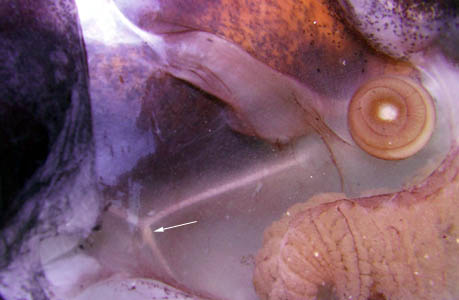
Figure. View of a portion of the mantle cavity of V. infernalis. The stellate ganglion (arrow), a characteristic structure of cephalopods, appears as only a slight enlargement of a large mantle nerve. The nerve running toward the oviducal gland is the fin nerve. Photograph by R. Young.


 Go to quick links
Go to quick search
Go to navigation for this section of the ToL site
Go to detailed links for the ToL site