Go to 2° Unipartite Upper Beaks
Go to 3° Unipartite Upper Beaks
Go to Unipartite Upper-Beak Survey
ORDER OEGOPSIDA:
OMMASTREPHIDAE: Eleven genera
UPPER BEAK CHARACTERISTICS
- Step: Absent.
- Shoulder Blade: Thick; occupies outer-most major layer of Shoulder but is partially covered laterally by thin layer of hyaline material.
- Shoulder Blade: Consists of two components (Inner and Outer). Inner Component forms separately from Outer Component and from pigmented Lateral Wall. Components fuse although line along fusion visible in cuts. Outer Component is continuous posteriorly with the Free Shoulder and the Inner Component extends posteriorly onto the Sessile Shoulder.
- Hyaline Matrix: Forms Inner (= medial) Part of Shoulder.
- Palatoshoulder Ridge: Absent.
- Pigmentation of posterior surface of Hyaline Matrix: During ontogeny pigmentation of Inner Component extends posteriorly onto lateral surface of Hyaline Matrix before pigmentation of posterior Lateral Wall reaches this point. This pigment is NOT continuous dorsally with that of the Bridge.
- Yellow line: Present. Lies between Shoulder Blade and Hood, may be difficult to see in heavily pigmented beaks.
Phylogenetic relationships.There is no compelling morphological data to determine familial relationships to the Ommastrephidae although Naef (1921-23) considered the Thysanoteuthidae to be their closest relative. The Bayesian analysis of Lindgren (2010) placed the Cranchiidae as the sister family to the Ommastrephidae with very good support; her Likelihood analysis placed the Ommastrephidae as sister to the Pyroteuthidae + Enoploteuthidae with weak support; her parsimony analysis has the family position of the Ommastrephidae unresolved. Lindgren et al. (2012) placed the Ommastrephidae as the sister family to the Thysanoteuthidae but with weak support.
Ommastrephidae: Structure
- EXTERNAL FEATURES
A. General Features
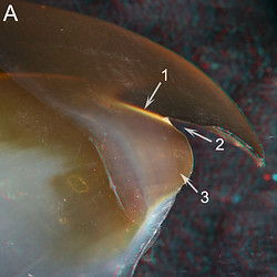

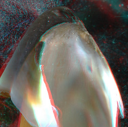
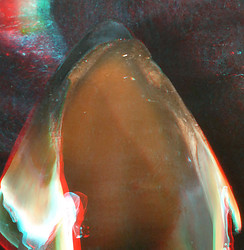
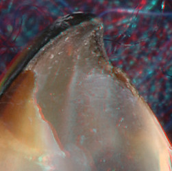
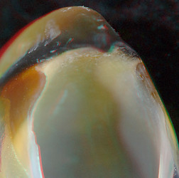
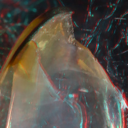
Sthenoteuthis oualaniensis, Mature female, 200 mm ML, 5.5 mm URL. A -Side view. B
- Oblique view.
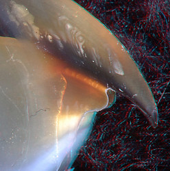
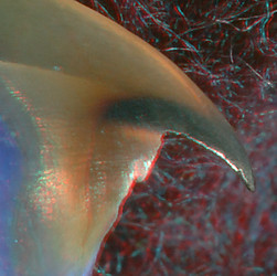
Nototodarus hawaiiensis, mature female,
130 mm ML, 4.2 mm URL. Oblique view.
ARROWS: 1 - Yellow Line. 2 - Jaw Angle. 3 - Outer Component of the Shoulder Blade. 4 - Palatine Ridges. 5 - Hyaline Matrix. 6 - Inner Component of the Shoulder Blade. 7 - Posterior end of fusion between Inner and Outer Components.
Fig. A - Shows the absence of a Step, the presence of a Yellow Line (1), an acute Jaw Angle (2) and a large Shoulder Blade (Outer Component) (3). Fig. B - Shows Palatine Ridges (4) characteristic of most Ommastrephinae which may or may not have a common origin with a Palatoshoulder Ridge but are not part of the Shoulder. Fig. C - Shows a member of the Todarodinae which lacks the Palatine Ridges. This particular beak has lost much of its pigment during preservation; the resulting transparency reveals the presence of a rather small Inner Component of the Shoulder Blade (6 on both Shoulders) as well as the junction of the Free and Sessile Shoulders (7) where the Components of the Shoulder Blade separate. The extent of the Hyaline Matrix (B, 5; C, 5) is difficult to see due to its translucency; look for small bits of dust that have settled on the surface of the Matrix (use magnified view - click on image).
B. Growth feature: Pigmentation of the posterior surface of the Hyaline Matrix.
 Click on an image to view larger version & data in a new window
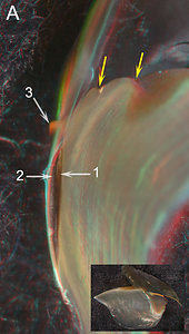
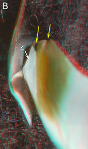
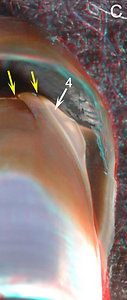
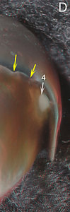
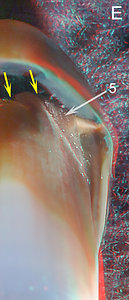
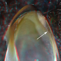
Figure. Sthenoteuthis oualaniensis. A - immature female, 144 mm ML, 4.3 mm URL. Posterior view. B - Female, 188 mm ML, 5.5 mm URL. Posterodorsal view. C. Mature male, 120 mm ML, 3.4 mm URL. D - Probable mature female, 196 mm ML, 5.3 mm URL Posterior view. E - Probable mature female, 200 mm ML, 5.8 mm URL. Posterior view.
ARROWS: 1 - Inner Component of the Shoulder Blade. 2 - Outer Component of the Shoulder Blade. 3 - Yellow Line. 4 -Hyaline Matrix. 5 - LW-B Continuum. Yellow arrows - Mark the two Palatine ridges of the Palate.These figures show the progressive pigmentation of the posterior region of the Shoulder. Unfortunately they are not all viewed from the same angle. Fig. A - The first area of the posterior Sessile Shoulder to be pigmented (1) is the extension of the Inner Component of the Shoulder Blade. Fig. B - The posterior pigmentation of the Lateral Wall has greatly increased but a broad region of Hyaline Matrix remains (4). As in the previous stage, there is no direct pigment connection between the Bridge and the Inner Component. However, at this stage, the posteroventral pigment of the Lateral Wall does barely connect with that of the Inner Component (out of the field of view in the image). Fig. C - The unpigmented Hyaline Matrix (4) has been reduced to a broad oval. Fig. D - The unpigmented Hyaline matrix has been reduced to a disc adjacent to the inner ridge of the Jaw-edge Extension. Fig. E - In this slightly larger female the posterior Shoulder is fully pigmented: continuous pigmentation exists between the Bridge, the Lateral Wall and the Inner Component of the Shoulder Blade which, here with an appropriate groove, seems to define a distinct LW-B Continuum (5).
- INTERNAL STRUCTURE: Longitudinal cuts through the Shoulder
With the Palatoshoulder Ridge virtually absent from the Shoulder, the Shoulder Blade is the only pigmented structure of the Shoulder during much of the beaks ontogeny. The Shoulder Blade, however, is large and complex, with an Inner Component and a thicker Outer Component. The two components separate posteriorly at the junction of the Free Shoulder and Sessile Shoulders.
Click on an image to view larger version & data in a new window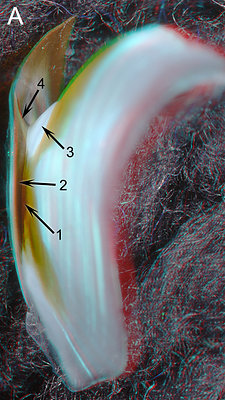
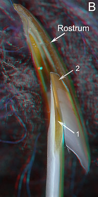
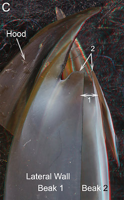
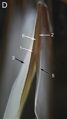
Figure. Sthenoteuthis oualaniensis, mature or nearly mature female, 188 mm ML, 5.5 mm URL. A - Posterior view. B-D - With longitudinal cut through shoulder. B - Oral-lateral view. C - Medial view of the cut shoulder superimposed on an oral view of the same beak. D - Photomicrograph (2D), oral view taken perpendicular to the plane of the cut.
ARROWS: 1 - Inner Component of the Shoulder Blade. 2 - Outer Component of the Shoulder Blade. 3 - Hyaline Matrix. 4 - Ridge along the medial side of the Jaw-edge Extension medially adjacent to the Yellow Line. 5 - Outer layer of hyaline material that covers the lateral surface of the Shoulder Blade. 6 - The line that separates the two components of the Shoulder Blade.Fig. A - The posterior view shows that the two pigmented components of the shoulder blade can be seen from this angle. The dark pigment of the Inner Component (1) merges with the lighter pigment of the Lateral Wall, and the Outer Component (2) continues on the Free Shoulder. Figs. B-D - The longitudinal cut through the Shoulder shows the Components more clearly. B - This oral-lateral view shows the tip of the Outer Component (2) lacks the outer cover of hyaline material. C - Two superimposed views of the same beak (medial view ("Beak 1") and oral view ("Beak 2")) allows a comparison of the Shoulder parts. The Inner Component (1) can often be recognized from a medial view when it is much smaller than the Outer Component. D - The higher resolution of this 2D image shows the structure of the beak in more detail. The thick layer of Hyaline Matrix (3) on the medial side of the shoulder thins anteriorly and barely extends over the leading edge of the Shoulder Blade. The thin external layer of hyaline material (5) thins anteriorly and is virtually absent near the anterior edge of the Blade.
- INTERNAL SRUCTURE: Cross-sectional cuts through the Shoulder
Click on an image to view larger version & data in a new window
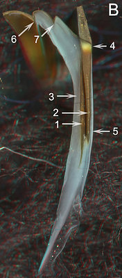
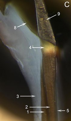
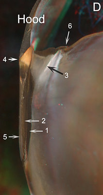
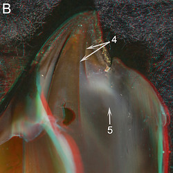
Figure. Same squid as above (188 mm ML, 5.5 mm URL). A - Posterolateral view. Insert shows position of cut in side view. B - Posterior view. C - Photomicrograph (2D), posterior view, taken perpendicular to plane of cut.
D - S. oualaniensis, mature female, 262 mm ML, 6.6 mm URL. Posterior view.
ARROWS: 1 - Inner Component of the Shoulder Blade. 2 - Outer Component of the Shoulder Blade. 3 - Hyaline Matrix. 4 - Yellow Line. 5 - Layer of hyaline material that covers the lateral surface of the Shoulder Blade. 6 - First ridge of Palate. 7 - Second ridge of the Palate. 8 - Bridge. 9 - Hood.
Fig. A - Shows the side and cut surface of the Yellow Line (4); a thin, lightly-pigmented layer covers the Yellow Line laterally. Fig. B - The cross-sectional cut shows the thick, pigmented Median Palate and the thin, pigmented Lateral Palate and the two prominent ridges of the palate (6, 7) characteristic of most members of the Ommastrephinae. How much of the latter constitutes the Bridge is unknown. One of these ridges appears, at first, to be a Palatioshoulder Ridge. But both could be new structures unrelated to the Ridge; neither has a Ridge-like role in the Shoulder. The plane of the cut is posterior to the fusion of the Components of the Shoulder Blade (1, 2). The thick Hyaline Matrix (3) covers the medial side of the Shoulder. Fig. C - A high resolution image of the central portion of Fig. B. The position of the Yellow Line (4) as a thick structure lying between the Outer Component of the Shoulder Blade and the Hood is clearly seen. Much of the hyaline material covering the lateral surface of the Shoulder Blade (5) appears dark due to reflection of light off the Blade and the thin external covering of the Yellow Line seen in Fig. A can barely be detected. Fig. D - A similar cut through the Shoulder of a much larger squid. Much of the Yellow Line (4) is covered Laterally by pigment of the Hood and Shoulder Blade. A thick core of Hyaline Matrix (3) still attaches to the inner side of the Yellow Line. Note that the tip of the black arrow also marks the inner edge of the cut surface.
- ORIGIN OF THE INNER COMPONENT OF THE SHOULDER BLADE.
Click on an image to view larger version & data in a new window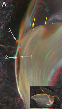
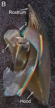

Figure. Sthenoteuthis oualaniensis, immature female. A - 144 mm ML, 4.3 mm URL. Posterior view; insert shows side and extent of pigmentation. B, C - 140 mm ML, 3.6 mm URL. Posterolateral view (B) and posterior-oral view (C). Most hyaline material has been dissolved, leaving pigmented layers and thin, white binding layers.
ARROWS: 1 - Inner Component of the Shoulder Blade. 2 - Outer Component of the Shoulder Blade. 3 - Yellow Line. 4 - Debris caught at the junction of the two Components. Yellow arrows - Mark the two ridges of the Palate.The Inner Component of the Shoulder Blade appears to have arisen by pigmentation of the lateral surface of the Hyaline Matrix or its associated "binding layer" rather than as an extension of pigment from the Bridge or posterior Lateral Wall. Fig. A - Shows that the Inner Component (1) is isolated from pigmented layers dorsally, posteriorly and ventrally. Figs. B, C - Show the same isolation in a beak that has lost most of the hyaline material via dissolution. (Surprisingly the thin, outer layer laterally covering the Shoulder Blade remains.) The Inner Component is isolated on all sides, except for its lateral fusion to the Outer Component, from any pigmented layer. On its medial side, the Inner Component is covered by a white, striated material which seems to be a "binding layer" to the Hyaline Matrix. This binding layer extends dorsolaterally beneath the Bridge and ventrally over the medial surfaces of the Inner and Outer Components. Fig. C - This more posterior view confirms the identification of the Inner Component by showing the fusion of the two Components (see 1, 2 on right side of image).
- CHANGES WITH GROWTH
- Sthenoteuthis oualaniensis:
Here we examine changes in the structure of the Shoulder Blade and the Hyaline Matrix.
Click on an image to view larger version & data in a new window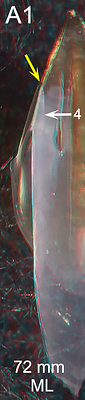

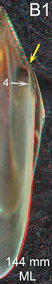
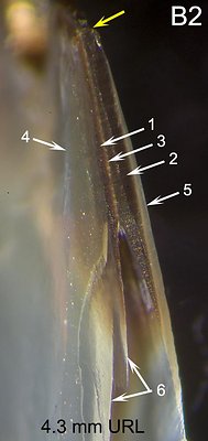
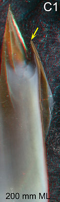

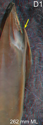
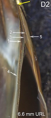

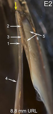
Figure. Sthenoteuthis oualaniensis with longitudinal cut through the shoulder. Images paired: oral view (3D) for viewer orientation on left and a higher resolution photomicrograph (2D) on the right. A - Immature, sex ?, 72 mm ML, 1.3 mm URL. B - Immature female, 144 mm ML, 4.3 mm URL. C - Mature female, 200 mm ML, 5.5 mm URL. D - Mature female, 262 mm ML, 6.6 mm URL. E - Mature female, 300 mm ML, 8.8 mm URL.
ARROWS: 1 - Inner Component of the Shoulder Blade. 2 - Outer Component of the Shoulder Blade. 3 - Line that separates the two Components. 4 - Hyaline Matrix. 5 - Layer of hyaline material covering external surface of Shoulder Blade. 6 (B2) - Indicates damage caused during the cut resulting in the posterior portion of the Inner Component of the Shoulder Blade breaking free from the Hyaline Matrix. Yellow arrows - Point to same spot on all beaks (anterior end of the Shoulder Blade where cut was made). - Nototodarus hawaiiensis (102 mm ML, 3.4 mm URL):
The following cuts were made to show that the features seen in Sthenoteuthis are representative of other Ommastrephids. Nototodarus is in a different subfamily from Sthenoteuthis and lacks the distinctive ridges of the Palate.
- Nototodarus hawaiiensis (160 mm ML, 5.3 mm URL): Click on an image to view larger version & data in a new window
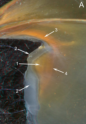
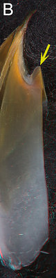
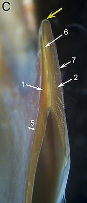
Figure. A - Medial view of the Shoulder region of the right half of the beak. B - Medial and slightly oral view of a beak fragment with a longitudinal cut of the Shoulder. C - Photomicrograph (2D), oral view, taken perpendicular to the cut surface of the Shoulder. ARROWS: Same as previous images of Nototodarus.
Fig. A - Comparsion of the two N. hawaiiensis of different sizes shows that, as expected, the relative size of the Inner Component (1) of the Shoulder Blade has increased greatly in the larger beak. In the larger beak much of the beak pigmentation has faded during preservation making some structures much easier to see. Fig. C - Surprisingly, the fusion of the two Components (6) does not appear as distinct a line as in Sthenoteuthis. The external layer of hyaline material (7), unlike Sthenoteuthis, extends to the anterior tip of the Shoulder Blade where it is continuous with the thin Hyaline Matrix from the medial side of the Shoulder. The Hyaline Matrix is thin anteriorly but not absent as in Sthenoteuthis but thickens on the posterior Shoulder as in Sthenoteuthis.
Fig. A - The Outer Component of the Shoulder Blade is well developed but the Inner Component has not yet appeared although a thick layer of opaque white material is present medial to the Shoulder Blade. Fig. B - The Inner Component has appeared but is much thinner than the Outer Component (don't be confused by the dark, medial side of the Inner Component that is visible through the Hyaline Matrix. The Hyaline Matrix is still thick. Fig. C - The Inner Component is much thicker and continuous with the posterior pigmented layer of the Lateral Wall. Anteriorly the Hyaline Matrix is greatly reduced in thickness. Figs. D, E - The trends continue and by Fig. E, both Components are about equal in thickness and the Hyaline Matrix is virtually absent from the area of the Shoulder.
Click on an image to view larger version & data in a new window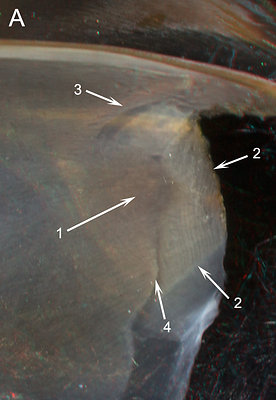
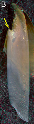
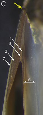
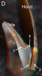
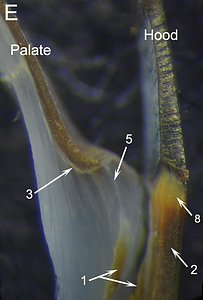
Figure. Nototodarus hawaiiensis. A - Medial view of the shoulder region of the left half of the beak. B - Oral-medial view of a beak fragment with a longitudinal cut of the shoulder. C - Photomicrograph (2D), oral view, taken perpendicular to the surface of the longitudinal cut of the Shoulder. D - Posterior view of a cross-sectional cut through the Shoulder. E - Photomicrograph (2D), posterior view of the same cross-sectional cut showing greater detail.
ARROWS: 1 - Inner Component of the Shoulder Blade. 2 - Outer Component of the Shoulder Blade. 3 - Rounded ridge. 4 - Posterior junction of the two Components of the Shoulder Blade (barely detectable). 5 - Hyaline Matrix. 6 - Midline between the two components of the Shoulder Blade. 7 - Layer of hyaline material of the external (= lateral) surface of the Shoulder Blade. 8 - Yellow Line. Yellow arrows - Point to same spot on both beaks (anterior end of the Shoulder Blade where cut was made).Fig. A - Medial view showing that the two components of the Shoulder Blade can sometimes be easily recognized without cutting the Shoulder. Fig. B - Overview of the cut beak for orientation. Fig. C. Detail of the cut shoulder showing structure very similar to that of Sthenoteuthis. The double ended arrow (5) indicates width of the Hyaline Matrix. The Inner Component of the Shoulder Blade (1) is in an early stage of development; posteriorly its thickness tapers to near extinction. Fig. D, E - These cuts show that the "rounded ridge" (A, 3; D, 3) is a simple fold of the pigmented Palate/Bridge that surrounds a lateral extension of the Hyaline Matrix and not a reduced Palatoshoulder Ridge. The Yellow Line (E, 8) has a similar structure to that of Sthenoteuthis. In Fig. E, the two parts of the beak fragment have broken slightly apart (1) although they had not broken in Fig. D.
- Sthenoteuthis oualaniensis:
THYSANOTEUTHIDAE: One species
UPPER BEAK CHARACTERISTICS
- Step: Absent.
- Shoulder Blade: Very thick, not covered lateraly by thin hyaline-like material.
- Shoulder Blade: Consists of two Components (Inner and Outer) separated by a distinct line in longitudinal section.
- Hyaline Matrix: Forms Inner (= medial) Part of Shoulder.
- Palatoshoulder Ridge: Absent.
- Pigmentation of posterior surface of Hyaline Matrix: During ontogeny pigmentation of Inner Component extends onto lateral surface of Hyaline Matrix before pigmentation of posterior Lateral Wall reaches this point. This pigment is continuous dorsally with that of the Bridge.
- Yellow Line: Present. Structure of Line different from that of ommastrephids.
Phylogenetic relationships. There is no compelling morphological data to determine familial relationships to the Thysanoteuthidae although Naef (1921-23) considered the Ommastrephidae to be their closest relative. In Lindgren's (2010) Parsimony analysis the family position of the Thysanoteuthidae is unresolved; in her Likelihood analysis the Thysanoteuthidae was placed as the basal family in the Oegopsida but with weak support; in her Bayesian analysis it was placed in the same position but with excellent support but its nearest relative appears well removed. In the Lindgren et al. (2012) analysis the Thysanoteuthidae was placed as the sister family to the Ommastrephidae but with weak support.
Thysanoteuthidae: Structure
- EXTERNAL FEATURES

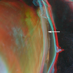
Thysanoteuthis rhombus, male, 310 mm ML, 4.4 mm URL. LEFT- Side view. LEFT MIDDLE - Oblique frontal view.
RIGHT MIDDLE - Posterior view, right half. Arrow - Inner Component of the Shoulder Blade.
T. rhombus, male, 735 mm ML, 6.7 mm
URL. RIGHT - Oblique-frontal view.At 310 mm ML, the absence of a Step, the acute Jaw Angle and the Yellow Line are apparent (Left); but the latter is not detectable in the beak of the 735 mm squid (Right). There is no indication of a Palatoshoulder Ridge in either beak although a slight ridge marking the position of the LW-B Continuum is present. In the smaller beak, the Lateral Wall is about 2/3 pigmented but the posterior surface of the Shoulder is still not fully pigmented as can be seen by a "window" on the beak's left side (Left, Middle). In the posterior view (Right Middle) the Inner Component (arrow) of the Shoulder Blade is separate from the more posterior pigment of the Lateral Wall by hyaline material (i.e., the "widow" seen in the previous image). This view also shows a clear connection between the pigment of the inner Component and that of the Bridge.
- INTERNAL STRUCTURE: Longitudinal Cuts
Click on an image to view larger version & data in a new window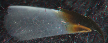

Figure. T. rhombus, 2.2 mm URL. TOP: Side view showing position of the longitudinal cut through the Shoulder. BOTTOM: - Oral view. Arrows: White - Outer Component of the Shoulder Blade. Black - Inner Component of the Shoulder Blade.
Bottom: In this small beak (2.2 mm URL) the pigmented Inner Component of the Shoulder Blade (black arrow) is distinct from posterior pigmentation of the Lateral Wall which is not present at this size (Top). While the longitudinal cut was not smooth, a line between the two pigmented components of the Shoulder Blade seems to be present. The medial Hyaline Matrix does not reach the anterior end the the Shoulder Blade and no hyaline material is present on the outer surface of the shoulder.
- INTERNAL STRUCTURE: Cross-sectional Cuts
Click on an image to view larger version & data in a new window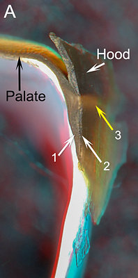
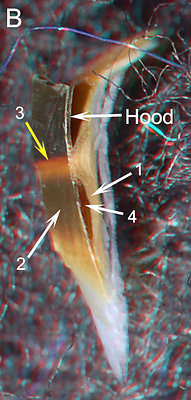
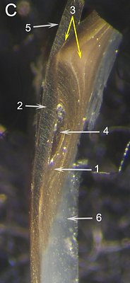
Figure. Thysanoteuthis rhombus, 2.2 mm URL, beaks from fish stomachs. A - Anterior view of a cut anterior to the Shoulder Pocket. B - Cut further posterior. Posterolateral View. C - Different beak, same size. Photomicrograph (2D), anterior view, perpendicular to plane of cut, shows more detail.
ARROWS: 1 - Inner Component of the Shoulder Blade. 2 - Outer Component of the Shoulder Blade. 3 - Yellow Line. 4 - Shoulder Pocket. 5 - Hood. 6 - Hyaline Matrix.
Figs. A, B - Show the relationship between the Yellow Line as seen on the outside of the Shoulder and in the cross-sectional cut of the Shoulder. Fig. A - In Thysanoteuthis, anterior to the Shoulder Pocket, the outer (=lateral) part of the Inner Component of the Shoulder is fused with the Outer Component but its inner (=medial) part appears to continue onto the Palate. Opposite the Yellow Line, the growth lines of the Inner Component are broadly separated. Fig C - Confirms this structure. The region of the Yellow Line has a "dome-like" appearance due, in part, to constriction of the Inner Component by the Shoulder Pocket. This "dome" has lighter color. At the top dome, where the Outer Component, the Inner Component and the Hood join, a thin, yellowish plate is present (3, left arrow). The lighter-colored areas of the dome and, particularly, the plate appear to be responsible for the color of the Yellow Line.
ORDER MYOPSIDA:
LOLIGINIDAE: 9 genera
UPPER BEAK CHARACTERISTICS
- Step: Absent.
- Shoulder Blade: Thick; occupies outer-most major layer of Shoulder but without lateral thin layer of hyaline material.
- Shoulder Blade: Consists of two Components (Inner and Outer) separated by distinct line in, at least, young beaks.
- Hyaline Matrix: Forms Inner (= medial) Part of Shoulder.
- Palatoshoulder Ridge: Absent.
- Pigmentation of posterior surface of Hyaline Matrix: During ontogeny pigmentation of Inner Component extends onto lateral surface of Hyaline Matrix before pigmentation of posterior Lateral Wall reaches this point. This pigment is continuous dorsally with that of the Bridge.
- Yellow line: Present. Weak in Sepioteuthis and Doryteuthis, absent in Uroteuthis. White Patch, apparently related to Yellow Line, present in Doryteuthis.
Phylogenetic relationships.The Loliginidae (or Myopsida when including the single species of the Australiteuthidae) has a long history of uncertainty in its phylogenetic position. Young et al. (1998), on morphological grounds, considered its position unresolved somewhere between typical sepioids and typical oegopsids. Numerous molecular studies have struggled with its position which has yet to receive a consensus. Lindgren (2012) places the five basal taxa of the Decapodiformes (Idiosepiidae, Sepiidae, Spirulida, Sepiolida, Loliginidae), which had unresolved relationships between them, in a clade which is sister to the Bathyteuthoidea + Oegopsida.
Loliginidae: Structure
- EXTERNAL FEATURES
We have only examined 3 of the 9 genera in the family; however, the 3 genera represent the 3 major clades within the family (Anderson, 2000, Pankey, et al., 2014). As in Ommastrephidae and Thysanoteuthidae a distinct but weak "yellow line" is present in Sepioteuthis (the sister genus to the rest of the family (Anderson, 2000) and becomes modified in (at least) one other genus (see below).
- Sepioteuthis
Sepioteuthis lessoniana, immature male, 64
mm ML, 1.0 mm URL. LEFT - Oblique view.S. lessoniana, female, 155 mm ML, 2.5 mm URL. MIDDLE - Side view. RIGHT - Frontal
view.The absence of a Step, the acute Jaw Angle (although slightly rounded at the Yellow Line) and the Yellow Line are apparent (Middle). No Palatoshoulder Ridge is seen (Left, Right). The two Components of the Shoulder Blade can be recognized in the frontal view of the left Shoulder (Right).
- Uroteuthis and Doryteuthis
The few beaks we have seen of species in these two genera, have a much more slender shoulder than S. lessoniana.
Uroteuthis sp., immature male, 46 mm ML,
1.0 mm URL. LEFT - Oblique view.Doryteuthis opalescens, off California, 128 mm ML, mature female. LEFT - Side view.
MIDDLE - Frontal view.The Shoulder has no Step (Middle). The Jaw Angle is often acute (Left, right Shoulder, Right, left Shoulder) but the more fragile structure of these beaks frequently results in damage to the anterior end of the Shoulder which can change the Jaw Angle. A weak Yellow Line is apparent in D. opalescence (Middle) but is not apparent in some specimens and was not apparent in Uroteuthis sp. There is no Palatoshoulder Ridge. However, a pigmented strip of the lateral wall seemingly terminates opposite the Shoulder (Right, arrow) as a low ridge; this strip, more anteriorly and opposite the White Patch, marks the junction of the Lateral Wall/Bridge with the Hood. Opposite the Shoulder Blade, however, the appearance of a Ridge results from the termination of the Hyaline Matrix (see below).
- Sepioteuthis
- INTERNAL STRUCTURE: Longitudinal Cuts
- Sepioteuthis Click on an image to view larger version & data in a new window
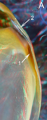
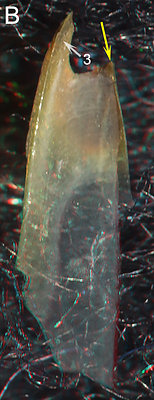
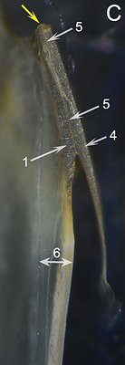
Figure. Sepioteuthis lessoniana, immature male, 64 mm ML, 1.0 mm URL. A - Posterior view. B - Medial view of left beak fragment. The beak shattered when cut and the hood is missing. C - Photomicrograph (2D), oral view taken perpendicular to the cut surface of the shoulder.
ARROWS: 1 - Inner Component of the Shoulder Blade. 2 - Hood (partially removed for better viewing). 3 - Palate (Hood broke off). 4 - Outer Component of the Shoulder Blade. 5 - Midline between the two components of the Shoulder Blade. 6 - Width of Lateral Wall, virtually all Hyaline Matrix. Yellow arrows - Point to same spot on beaks B and C, and mark the anterior end of the Shoulder Blade where cut was made.Fig. A - The posterior view of the beak S.lessoniana shows a pigmented region on the Sessile Shoulder (1) corresponding to the Inner Component of the Shoulder Blade. A lighter line of debris just beyond the point of the arrow indicates the junction of the Free and Sessile Shoulders. Fig. C - The photomicrograph of the longitudinal cut shows a distinct line been the Inner and Outer Components of the Shoulder Blade (5) (this line is interrupted by an artifact of an imperfect cut). The thick Hyaline Matrix extends nearly to the anterior end of the Shoulder Blade. No hyaline layer appears to be present on the lateral (ie, outer) side of the Shoulder Blade.
- Uroteuthis and Doryteuthis Click on an image to view larger version & data in a new window
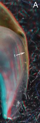
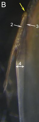

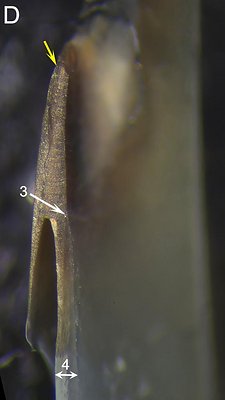
Figure. Uroteuthis sp. (A, B) and D. opalescens (C, D), (same beaks as above: External Features). A (Uroteuthis sp.) - Posterior view of right side of the beak. B - Photomicrograph (2D), oral view perpendicular to cut surface of Shoulder. C (D. opalescens) - Oral-medial view of the beak with a longitudinal cut through the shoulder (3D). D - Photomicrograph (2D) oral view perpendicular to cut surface of Shoulder.
ARROWS: 1 - Inner Component of the Shoulder Blade. 2 - Midline between the two Components of the Shoulder Blade. 3 - Anterior limit of the Hyaline Matrix. 4 - Width of the Hyaline Matrix. 5 - Pigment strip (see below). 6 - Palate. Yellow arrows - Point to same spot on all beaks (anterior end of the Shoulder Blade where cut was made).
Fig. A - The Inner Component of the Shoulder Blade (1) in this young Uroteuthis beak is similar to that seen in Sepioteuthis and is continuous with the pigment of the Bridge dorsally. Fig. B - The Inner Component barely extends posteriorly beyond its junction with the Outer Component but extends anteriorly to the tip of the Shoulder Blade. The thick Hyaline Matrix (4) medial to the Shoulder Blade, thins anteriorly and ends (3) short of the anterior tip of the Shoulder Blade. A line (2) separates the two Components of the Shoulder Blade. Fig. C - A pigmented strip (5) in this mature Doryteuthis marks the fusion of the Palate to the Hood. This pigment only seems to continue, less well defined onto the Shoulder because of the anterior edge of the Hyaline Matrix (3) which is much further posterior than in the young Uroteuthis beak. Fig. D - The pigmentation of the Inner Component posterior to the junction with the Outer Component shows a gradation from darker anteriorly to lighter at its posterior termination which approximately mimics the more anterior pigment strip. Superimposed on this gradient is the anterior termination of the Hyaline Matrix (3) at the start of the gradient. Therefore what appears as a possible Palatoshoulder Ridge, (see Right image in External Features above) is not a Ridge. A thin line separating the two pigmented parts of the shoulder blade is not seen, possibly as a result of age.
- Sepioteuthis
- INTERNAL STRUCTURE: Cross-sectional Cuts
- Sepioteuthis Click on an image to view larger version & data in a new window
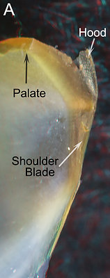
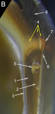
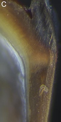
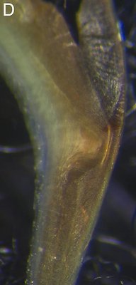
Figure. Sepioteuthis lessoniana, 155 mm ML, 2.5 mm URL. A - Anteror view of the cut Shoulder (3D), most of the Hood was lost during the cut. B-D - Photomicrographs (2D) showing greater detail. B - Anterior view with slight lateral tilt. C - A slightly deeper "cut" due to sanding. View perpendicular to the plane of the cut and showing better detail. D - A slightly deeper "cut" than C, same view; accidental focus beneath the cut surface showing surprising amount of detail.
ARROWS: 1 - Inner Component of the Shoulder Blade. 2 - Outer Component of the Shoulder Blade. 3 -Hyaline Matrix. 4 - Yellow Line structures, right two arrows indicate "plates", left arrow indicates "dome". 5 - Preparation artifact. 6 - Outer (= lateral) edge of cut surface. 7 - Yellow Line (withreddish tint) on lateral surface of beak.Fig. A - This 3D view is an aid in viewer orientation. Fig. B - The cut was not smooth so the beak was embedded in plastic and sanded but the technique resulted in some lack of clarity. The slight tilt in this photograph allows the viewer to see the position of the Yellow Line on the outer surface of the beak when examining the internal structure. Note the position of the outer edge of the cut (6); everything to the right of this is on the outside of the beak. Fig. C - This view gave the best detail of the cut surface. Fig. D - A missed focus slightly below the cut surface shows surprising, but not well understood, detail.
In Sepioteuthis, as in Thysanoteuthis, Inner component of the Shoulder Blade (B, 1) is mostly responsible for formation of the "dome" at the site of the Yellow Line; its growth lines, with greater separation laterally, can be seen entering and leaving the dome (B, 4, C). This "dome" has lighter color. At the top dome, where the Outer Component, the Inner Component and the Hood approximately join, two thin, lightly-colored plates are present (B, 4, C and D). The dome and, particularly, the plates appear to be responsible for the color of the Yellow Line. - Doryteuthis
Click on an image to view larger version & data in a new window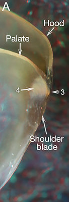
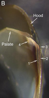
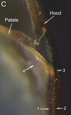
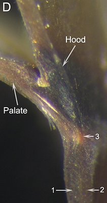
Figure. D. opalescens, mature female, 128 mm ML, 1.4 mm URL. A. 3D image of the cut beak. B-D. Photomicrographs (2D) at slightly different angles and magnification.
ARROWS: 1 - Inner Component of the Shoulder Blade. 2 - Outer Component of the Shoulder Blade. 3 - Yellow Line. 4 -White Patch.
Fig. A - The cut seen here was not smooth and subsequent sanding of the cut surface was necessary for higher resolution photographs. This image shows the structure that creates the Yellow Line (3) and and what appears to be its modification, the White Patch (4), lying over the oral surface of the palate in the region of the Jaw Edge Extension. The White Patch (4) is also seen in Figs. B and C and recalls a similar appearing region in most sepiolids. Fig. D - Shows the most detailed view of the structure in the region of the Yellow Line (3).
In spite of the much thinner upper Shoulder in Doryteuthis, its structure appears to be similar to that of Sepioteuthis. The Inner Component of the Shoulder Blade, however, forms a more flattened "dome" (B, D) with less well-defined structure with a possible "plate," at its apex. The dome is covered laterally by a thicker more heavily pigmented layer than in Sepioteuthis that, apparently, is formed by the fusion of the hood and Outer Component of the Shoulder Blade (B, C). This layer would greatly reduce the visibility of the Yellow Line from the exterior.
ORDER OEGOPSIDA:
ARCHITEUTHIDAE:One species
We have not had the opportunity to cut any Architeuthis beaks; we have examined only four large beaks and none of intermediate size. Therefore its inclusion as a 1° Unipartite beak is uncertain.
UPPER BEAK CHARACTERISTICS
- Step: Absent.
- Shoulder Blade: The "outer component" of the Shoulder Blade has a thin free-edge with pieces broken off, similar to many Bipartite beaks.
- Shoulder Blade: One beak showed a possible Inner Component of the Shoulder Blade (Middle, arrow) in a medial view of the Shoulder.
- Hyaline Matrix: Forms Inner (= medial) Part of Shoulder.
- Palatoshoulder Ridge: None recognizable.
- Pigmentation of posterior surface of Hyaline Matrix: Only one beak had incompletely pigmented the posterior Shoulder. It showed a possible posterior extension of an Inner Component on the Shoulder Blade onto the posterior Hyaline Matrix from the junction of the Sessile and Free Shoulders.
- Yellow Line: Present. Structure unknown.
Phylogenetic relationships. On morphological grounds, the Architeuthidae is placed next to the Neoteuthidae (Roper and Young, 1972) in a clade including the enoploteuthid and histioteuthid families. All four molecular analyses by Lindgren (2010) and Lindgren et al. (2012) strongly confirm the sister relationship of the Architeuthidae and Neoteuthidae. In addition, Lindgren (2010) in her Likelihood and Bayesian analyses found the Architeuthidae + Neoteuthidae clade sister to the histioteuthid families with excellent support. But the Lindgren et al. (2012) analysis did not show the latter relationship.
Architeuthidae: External features
Architeuthis sp., male, 1.1 m ML. LEFT - Side view . MIDDLE - Oblique-oral view.
Architeuthis sp., immature female, 1.2 m
ML, 16 mm URL. RIGHT - Posterior view.
The absence of a Step and the presence of a Yellow Line are apparent (Left, Middle). The Jaw Angle is generally acute but easily altered by damage. A possible Inner Component of the Shoulder Blade can be seen through the medial Hyaline Matrix in one beak (Middle, arrow) and a possible posterior extension of a Inner Component can be seen on another (Right, arrow) but seems less well-defined than is typical of 1° Unipartite beaks.
COMMENTS ON 1° UNIPARTITE BEAKS (except Architeuthis):
The Shoulders of Thysanoteuthis and loliginids examined show considerable similarities. Although we don't know how the Inner Component of the Shoulder Blade forms in these two taxa, they are similar in having a continuous pigmented layer that extends from the Inner Component to the Bridge while being separated from posterior Lateral Wall pigment. This occurs over a range of sizes (i.e., at least 70-310 mm ML in Thysanoteuthis and 38-? mm ML in Sepioteuthis). A similar pigment strip is not found in ommastrephids we examined although all three taxa share a temporary gap in pigmentation between the Inner Component the posterior Lateral Wall pigment. The structure that is responsible for the Yellow Line in Thysanoteuthis and the loliginids is similar but appears to be very different from that of ommastrephids.
The similarities in the upper beak between the Thysanoteuthidae and Loliginidae suggest that they are phylogenetically closely related. No recent morphological or molecular data support this relationship although Lindgren 2010 placed Thysanoteuthis at the base of the Oegopsida (in two of three analyses) with all other oegopsids as its sister group. The phylogenetic position of the Loliginids is still debated but it, at best, it is separated from the base of the Oegopsida by the Bathyteuthoidea.
- Sepioteuthis


