Go to 1° Unipartite Upper Beaks
Go to 2° Unipartite Upper Beaks
Go to Unipartite Upper-Beak Survey
On this page we include taxa with Unipartite beaks that have the two essential features of a Unipartite beak(i.e., no Step, absence or virtual absence of a Palatoshoulder Ridge) but lack some of the other Unipartite-beak features. All of these taxa have close relatives whose beaks are either Bipartite or Tripartite. The taxa included here are all oegopsids: Brachioteuthidae (one genus), Joubiniteuthidae, Cranchiinae (all genera), Pyroteuthidae and Lampadioteuthinae.
ORDER OEGOPSIDA:
BRACHIOTEUTHIDAE: Slosarczykovia circumantarctica
UPPER BEAK CHARACTERISTICS:
- Step: Absent.
- Shoulder Blade: Somewhat thick. Occupies outer-most major layer of Shoulder; covered laterally by extremely thin layer of hyaline material.
- Shoulder Blade: Consists of two Components (Inner and Outer). Inner Component forms from pigmented Lateral Wall. A line separating the Components present early but not in well-pigmented beaks.
- Hyaline Matrix: Forms Inner (= medial) Part of Shoulder.
- Palatoshoulder Ridge: None recognizable.
- Pigmentation of posterior surface of Hyaline Matrix: Ventral region of Matrix covered via intrusion of a Shoulder Pocket with Lateral-Wall pigment (extension of latter forms Inner Component of Shoulder Blade).
- Yellow Line: Absent; White Patch absent.
Phylogenetic relationships. Morphology has not provided convincing evidence of relationships. On the basis of molecular work, Lindgren et al. (2012) found the family sister to the Gonatidae. Lindgren (2010) found the relationships undefined in her Parsimony analysis but placed the family as a sister group to the Cycloteuthidae in both her Likelihood and Bayesian analyses with weak support in the former but with strong support in the latter.
Brachioteuthidae: Structure
- EXTERNAL FEATURES
We have seen two species of Brachioteuthidae, Slosarczykovia circumantarctica and Brachioteuthis beanii. The latter is not treated here as it has a Type 3 Bipartite beak; the former is treated below although we had only one beak to examine.
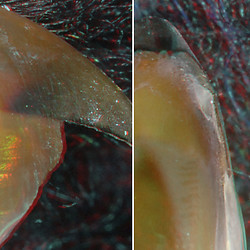
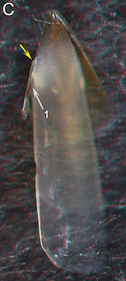
Slosarczykovia circumantarctica, female,
115 mm ML, 2.5 mm URL. LEFT - Side view,
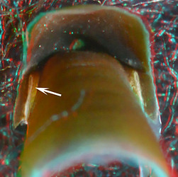
RIGHT - Front-oral view.S. circumantarctica, male, 67 mm ML, 1.4 mm URL. LEFT - Oblique view. RIGHT - Posterior
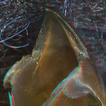
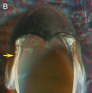
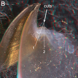
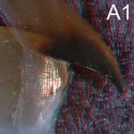
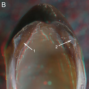
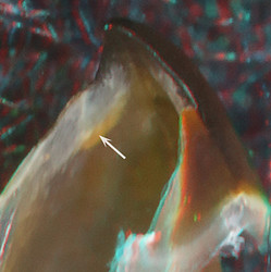
view.The larger, heavily-pigmented beak (Left pair) shows no Step, no Yellow Line, and has an acute Jaw Angle; its front-oral view (Left middle) shows a Shoulder mostly composed of a thick, complex Shoulder Blade although, at this late stage, the Inner Component may be combined with pigmentation of the Hyaline Matrix. In the smaller beak of S. circumantarctica (Right pair) the Lateral Wall is about two thirds pigmented which is sufficient pigmentation for a Palatoshoulder Ridge to be present if it were to occur, but there is no trace of one. A Shoulder Pocket (1, 2) enables pigment of the outer layer of the Lateral Wall to extend over the lateral surface of the Hyaline Matrix and onto to the Shoulder Blade (see cut below) where it becomes the Inner Component of the Shoulder Blade. This positioning of the outer pigmented layer of the Lateral Wall anteriorly isolates a thick pocket of Hyaline Matrix (Left middle, 3 Right, 3).
- INTERNAL SRUCTURE: Longitudinal cuts through the Shoulder.
 Click on an image to view larger version & data in a new window
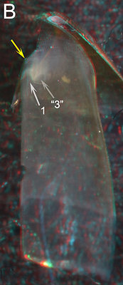
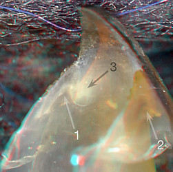
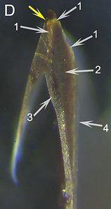
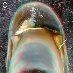
S. circumantarctica, 67 mm ML, 1.4 mm URL. A - Side view showing the position of the cut. B & C - Medial and Oral views to aid the viewer in orientation. D - Photomicrograph (2D), oral view taken perpendicular to the cut.
ARROWS - 1 - Pigmented Shoulder Pocket. 2 - Hyaline Matrix. "3" - Unpigmented dixc-shaped, unpigmented, posterior surface of Hyaline Matrix (see Right image in table above). 3 - Outer Component of the Shoulder Blade. 4 - Break, formed during the cut, in the outer layer of pigment in the Lateral Wall. 5 - Extension of the Shoulder-Pocket pigment onto the Outer Component of the Shoulder Blade as the Inner Component.
The Inner Component of the Shoulder Blade is formed by the anterior extension of pigment from the Shoulder Pocket (D, 5) which, in turn, is from the outer pigmented Layer of the Lateral Wall (D, 4) (note that the Lateral Wall has a thick inner unpigmented layer of hyaline material). The Outer Component is covered laterally by a very thin layer of hyaline material (D), Both S. circumantarctica and species of the Cranchiinae (see below) appear to use the same approach in obtaining a Unipartite beak.
JOUBINITEUTHIDAE: single known species
UPPER BEAK CHARACTERISTICS
xxx
- Step: Absent.
- Shoulder Blade: Generally thin. Occupies outer-most major layer of Shoulder; not covered laterally by thin layer of hyaline material.
- Shoulder Blade: Consists of one component.
- Hyaline Matrix: Forms Inner (= medial) Part of Shoulder.
- Palatoshoulder Ridge: Virtually absent; rudiment present.
- Pigmentation of posterior surface of Hyaline Matrix: Mostly from pigment arising at medial side of Hyaline Matrix in a typical Bipartite manner.
- Yellow Line: Absent; White Patch absent.
Phylogenetic relationships. The Joubiniteuthidae is one of the chiroteuthid families (Tree of Life), perhaps most closely related to the Mastigoteuthidae (Lindgren, 2010). Species of these families mostly have Bipartite beaks.
Joubiniteuthidae: Structure
- EXTERNAL FEATURES
We examined four beaks of Joubiniteuthis portieri and cut two of them.
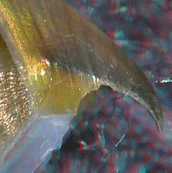

J. portieri, male ? (i.e., well pigmented at small size), 42 mm ML, 1.0 mm URL. LEFT - Side view. MIDDLE - Oblique view. RIGHT -
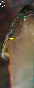
Posterior view.The Shoulder has no Step or Yellow Line (Left). The Jaw Angle varies from oblique to acute generally depending on beak size. The posterior pigmentation of the Lateral Wall mostly parallels the front of the Lateral Wall and Shoulder (Left, Middle) rather than crossing over it on the medial side to form a Palatoshoulder Ridge; instead, it terminates at the LW-B Continuum (Right, arrow) which pigments the back of the Shoulder in a manner typical of a Bipartite beak. A possible Palatoshoulder Ridge (Middle, arrow), that is more prominent in this beak than generally seen, is more a remnant than a Ridge (see below).
- INTERNAL SRUCTURE: Longitudinal cuts through the Shoulder.
- Small beak (1.0 mm URL) about 3/4 pigmented.
Click on an image to view larger version & data in a new window
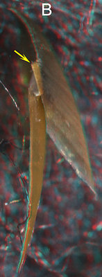

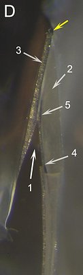
Figure. J. portieri, 42 mm ML, 1.0 mm URL. A: Side view of the cut beak showing where the longitudinal cut was made. B - Oral-lateral view. C - Oral view. D - Photomicrograph (2D), oral view taken perpendicular to the cut surface.
ARROWS: C1 - Remnant of Palatoshoulder Ridge. D1 - Approximate spot where a series of irregular ridges form a remnant of a blunt Palatoshoulder Ridge. 2 - Hyaline Matrix. 3 - Pigment covering much of the Posterior surface of the Hyaline Matrix. The space between the two arrows is not yet pigmented (compare to RIGHT figure in above table). Yellow arrows - All point to the same spot on the beak.The "Palatoshoulder Ridge" is not a simple ridge but a series of thick and thin ridges that intersect the cut (A, 1) where they are barely recognizable from one another, and which, at best, together form only a remnant of a true Palatoshoulder Ridge. The appearance of the ridges varies among specimens.
- Large beak (1.8 mm URL) near full pigmentation.

J. portieri, sex ?, 85 mm ML, 1.8 mm URL. A - Side view (top), oblique view (bottom). B - Front view. C - Oral-front view. D -
Photomicrograph (2D) of the cut shoulder taken perpendicular to the plane of the cut.ARROWS: 1 - Whitish/yellowish remnant of the Hyaline Matrix. 2 - Former Hyaline Matrix now pigmented. 3 - Pigmented posterior surface of the Hyaline Matrix. 4 - Hyaline material. Yellow arrows - Indicate same point on all beaks.
Figs. A and B show the beak before it was cut; note the acute Jaw Angle. In B two heavily pigmented layers converge at the anterior apex of the Shoulder separated by a white layer dorsally and ventrally. The left Shoulder, especially, present a view similar to a more typical Unipartite beak. The dorsal white layer appears to represent the tip of a thick Hyaline Matrix (C, 1) is actually a very thin remnant of the Matrix (D, 1). The nearly complete posterior pigmentation of the Matrix seen the smaller beak above is complete in this larger beak (3) and this pigmentation can be recognized as different from the general pigmentation of the Hyaline Matrix (2). Note that the angle at the junction of the Free and Sessile Shoulders has changed dramatically between the two beaks and that a typical layer of clear hyaline material covers much of the oral surface of the Shoulder and extends posteriorly over much/all of the Lateral Wall.
- Small beak (1.0 mm URL) about 3/4 pigmented.
Comments:
The typical Bipartite pigmentation of the posterior Shoulder seen here in this Unipartite beak clearly indicates the Bipartite origin of this beak. The Unipartite appearance of the beak seen in Fig. B appears to have developed simply through the pigmentation of the Hyaline Matrix. While this figure looks similar to the front-oral view of Slosarczykovia (above) the means used to obtain this condition seems to be entirely different (however, see below). Small Joubiniteuthis beaks tend to have a slightly oblique Jaw Angle while large beaks (i.e., 1.8 and 2.2 mm URL) have a distinctly acute Jaw Angle. Perhaps the late ontogenetic shape change is related to their incomplete transition to a Unipartite beak.
CRANCHIIDAE: CRANCHIINAE: Cranchia, Liocranchia and Leachia
UPPER BEAK CHARACTERISTICS
- Step: Absent.
- Shoulder Blade: Thin; outermost layer of Shoulder; without partial cover by thin layer of hyaline material.
- Shoulder blade: Consists of two (?, see below) Components. No central "line" separating components, in longitudinal section, in well-pigmented beaks.
- Hyaline Matrix: Forms Inner (= medial) Part of Shoulder.
- Palatoshoulder Ridge: Absent.
- Pigmentation of posterior surface of Hyaline Matrix: Pigmentation begins as in Bipartite beaks but intrusion of a Shoulder Pocket with Lateral-Wall pigment covers ventral region of Matrix early-on.
- Yellow Line: Absent; White Patch absent.
Phylogenetic relationships: There is no convincing evidence for family relationships based on morphology. Lindgren (2010) in a Bayesian analysis of molecular data, found the Cranchiidae to be a sister group of the Ommastrephidae with strong support but her other two analyses found no clear family relationships nor did Lindgren, et al. (2012).
Cranchiinae: Structure
- EXTERNAL FEATURES
We examined five beaks of Liocranchia reinhardti and cut two , only one of which is close to a critical size for determining structure.

Liocranchia reinhardti, sex ?, 100 mm ML, 1.4 mm URL. A - Side view. B - Medial view
of left half of beak showing where cuts were made to examine shoulder structure.
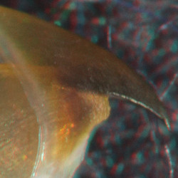
Cranchia scabra, immature female, 127 mm
ML, 2.6 mm URL. Posterior view.A Step and a Yellow Line are absent (A). When the Shoulder is not damaged a Tooth is generally present (B), that generally accentuates with growth, and gives the beak an acute Jaw Angle. The medial view of a half beak shows where the cuts (seen below) were made. A posterior view (C) shows the early pigmentation of the posterior surface of the Hyaline Matrix that looks much like that of a Bipartite beak but this changes quickly with the development of a Shoulder Pocket (see below).
- INTERNAL SRUCTURE: Longitudinal cuts through the Shoulder.
- Relatively small beak: 1.4 mm URL.
We examine two cuts through the shoulder of this beak. Cut 1 passes through the Shoulder Blade and the dorsal, thick region of the Hyaline Matrix. Cut 2 passes through the Shoulder Blade and the Shoulder Pocket that carries the pigmented Lateral Wall (see Fig. B above).
Click on an image to view larger version & data in a new window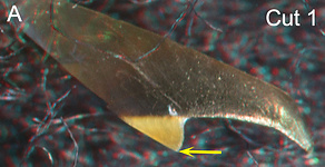
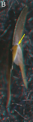

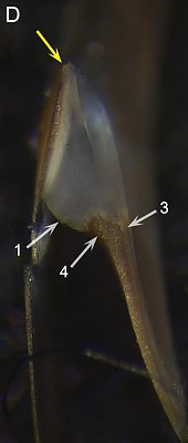
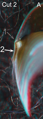
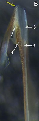
Figure. L. reinhardti, 100 mm ML, 1.4 mm URL. Cut 1 (A-D): A- Side view of of beak fragment showing where Cut 1 was made. B - Oral-lateral view of Cut 1. C - Oral view of Cut 1. B and C orient viewer. D - Photomicrograph (2D), oral-posterior view taken perpendicular to the cut surface. Cut 2 (A, B): A - Posterior view showing angles of cuts. B - Photomicrograph (2D), oral view taken perpendicular to plane of the cut.
ARROWS: Cut 1: 1 - Unpigmented posterior end Hyaline Matrix. 2 - Possible remnant of the Palatoshoulder Ridge dorsal to cut surface. 3 - Pigmented layer corresponding to the possible Palatoshoulder Ridge remnant seen in 2. Yellow arrows - indicate same point in all four images of Cut 1, and close to this point in Cut 2B.
Cut 2: A, 1 - Indicates direction of cut 1. A, 2 - Indicates direction of Cut 2. B, 1 - Reduced unpigmented posterior end of the Hyaline Matrix. B, 3 - Remnants (?) of the Palatoshoulder Ridge buried within the Hyaline Matrix. B, 4 - Lateral Wall pigment of Shoulder Pocket. B, 5 - Hyaline Matrix.
In Cut 1, D pigment continuous with the posterior Lateral Wall pigment has only slightly encroached onto the posterior surface of the Hyaline Matrix from the latter's medial side. In Cut 2, B this pigmented layer is much longer as the pigment of the Shoulder Pocket covers much of the posterior surface of the Hyaline Matrix. This pigment has nearly reached the Shoulder Blade and appears to be in an early stage of filling this gap (note the white area of Hyaline Matrix plus the yellow-brown streaks) and forming a Inner Component to the Shoulder Blade. The Palatoshoulder Ridge is virtually absent with only possible irregular remnants seen in the cuts (Cut 1 - D3; Cut 2 - B3). The formation of a Shoulder Pocket closes over the ventral region of the Hyaline Matrix but leaves a thick pocket of Matrix dorsally (2, A).
- Relatively large and more heavily pigmented beak: 1.9 mm URL.


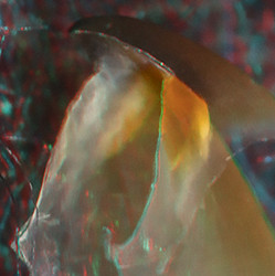
L. reinhardti, male, 180 mm ML, 1.9 mm URL. A1 - Side view. A2 - Oblique view. B - Front-oral view. C - Oral-front view.
D - Photomicrograph (2D) of the cut shoulder taken perpendicular to the cut surface.
ARROWS: 1 - Thick, rounded, pigmented ridge extending from the Shoulder onto the Palate. 2 - Jaw-angle Pocket. 3 - Small wedge of Hyaline Matrix residue. 4 - A thin layer of hyaline material along the medial side of the Shoulder that thickens as it extends posterior to the Shoulder. Yellow arrows: Point to the same spot (anterior end of cut) on Figs. C and D to aid viewer orientation.
The beak has a broad pigmented ridge (1) that extends from the Shoulder and the Palate. The ridge seems to have been formed mostly by pigmentation of the Hyaline Matrix (D1). A broad Jaw-angle Pocket is present (2) covered by, apparently, a thin residue of the Hyaline Matrix which extends (3) along the anterior edge of the pigmented Shoulder.
Comments.
There is strong similarity in the structure of the Shoulder of the larger Liocranchia reinhardti and the larger Joubiniteuthis joubini (Fig. D in both species). These beaks have arrived at a similar structure but from seemingly different paths. Also there is strong similarity in the younger Slosarczykovia circumantarctica and the younger L. reinhardti which have taken similar paths. It would be interesting to know the structure of a large beak of the former. Note the similarity between the Shoulders of the large beaks of these three species:
Click on an image to view larger version & data in a new window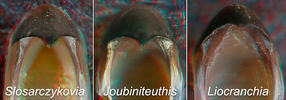
- Relatively small beak: 1.4 mm URL.
PYROTEUTHIDAE: 2 genera
UPPER BEAK CHARACTERISTICS
- Step: Absent.
- Shoulder Blade: On outer major part of Shoulder; without partial cover by thin layer of hyaline material.
- Shoulder Blade: Consists of one Component with possible late development of an Inner Component. No central "line" is apparent, in longitudinal section.
- Hyaline Matrix: Forms Inner (= medial) Part of Shoulder, covers most of medial remnant of Palatosholder Ridge.
- Palatoshoulder Ridge: Distinct remnant present, mostly buried within the Hyaline Matrix; composed, apparently, of several irregular layers with poorly defined boundries.
- Pigmentation of posterior surface of Hyaline Matrix: Unknown
- Yellow Line: Absent; White Patch absent.
Phylogenetic relationships: The Pyroteuthidae is placed in the enoploteuthid families (Ancistrocheiridae, Enoploteuthidae, Pyroteuthidae and Lycoteuthidae) on the Tree of Life. There is reasonable morphological support for this grouping (Young and Harman, 1998) and especially the close relationship between the Pyroteuthidae and the Lycoteuthidae (Herring, et al., 1985). Molecular support for the group (without Ancistrocheiridae due to lack of data) has been mixed. Lindgren's (2010) Bayesian analysis placed the three families together with strong support; her Maximum-likelihood analysis placed Pyroteuthidae and Enoploteuthidae together with good support, as did Lindgren et al. (2012) but did not place Lycoteuthidae nearby; her (2010) parsimony analysis had relationships of the three undefined.
Pyroteuthidae: Structure
- EXTERNAL FEATURES
A. General features.
We have examined six beaks of Pyroteuthis. addolux. The shoulder structure shows virtually no similarity to the Tripartite beaks of its close relatives among the Enoploteuthidae and Lycoteuthidae. The beaks are small and difficult to examine and conclusions are tentative.
Pyroteuthis addolux, immature female, 31
mm ML, 0.8 mm URL. Oblique view.
P. addolux, mature female, 38 mm ML, 0.7 mm URL. MIDDLE - Side view. RIGHT - Oblique
view.The beak has no Step and no Yellow Line and has an acute Jaw Angle (Middle). A distinct remnant of the Palatoshoulder Ridge (Right, arrow) remains. The Ridge has lighter pigmentation due to scattered back-light from the Hyaline Matrix. The Ridge remnant is similarly visible in the Left image and many of the images below.
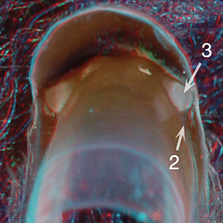
Posterior views of the three beaks to show pigmentation of the posterior surface of the Hyaline Matrix.
B. Posterior pigmentation of the Hyaline Matrix.
Click on an image to view larger version & data in a new window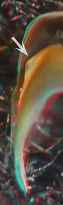
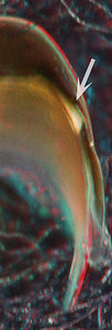
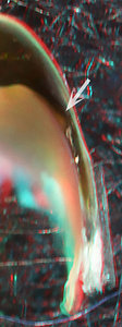
Figure. Posterior views of one side of each of the three beaks. Left - 31 mm ML, Middle - 33 mm ML. Right - 38 mm ML.
The beak of the 31 mm squid (left) shows a large unpigmented area (arrow) along the posterior end of the Sessile Shoulder. In the 33 mm-squid beak (middle) the unpigmented region is much smaller and concentrated near the Jaw-edge Extension, as is typical of Unipartite beaks at this stage of pigmentation. This area is more opaque white than in the 31mm-squid beak. In the 38 mm-squid beak (right), the posterior pigmentation is virtually complete (the white area below the pigmented bridge is debris from incomplete cleaning).
- INTERNAL STRUCTURE: Longitudinal cuts through the Shoulder of: (1) an immature female; (2) a just-mature female; (3) a large-mature female.
Click on an image to view larger version & data in a new window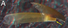
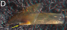
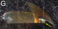
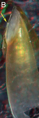
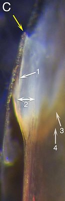
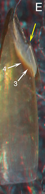
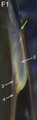
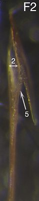
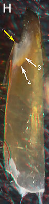
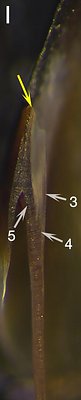
Figure. The same Pyroteuthis addolux beaks, with longitudinal cuts through the shoulders. A, D, G - Side view of beak fragments showing where the cut was made (Yellow arrow). A-C - Immature female, 31 mm ML, 0.8 mm URL. D-F2 - Just mature female, 33 mm ML, 1.2 mm URL. G-I - Larger mature female, 38 mm ML, 0.7 mm URL. B, E, H- Oral views of entire beak fragment to aid in orientation. C, F1, F2, I - Photomicrographs (2D) of the cut shoulder, oral view taken perpendicular to the plane of the cut.
ARROWS: 1 - Inner Component (?) of Shoulder Blade. 2 - Hyaline Matrix. 3 - Remnant of the Palatoshoulder Ridge. 4 - Base of the Palatoshoulder Ridge remnant. 5 - Full pigmentation of posterior Hyaline Matrix. Yellow arrows: Point to the same spot on all beaks.In the 31 mm ML, immature female (A-C), a short "spur" (1) extends posteriorly off the Shoulder Blade and onto the unpigmented surface of the Hyaline Matrix; a thin short pigment strand also extends anteriorly off the pigmented Lateral Wall onto the Hyaline Matrix (no arrow). These pigmented bits may be the initial step in the pigmentation of the posterior surface of the Hyaline Matrix, or the "spur" could be the initial formation of the Inner Component of the Shoulder Blade. Or, both options may be true.
The heavily pigmented Lateral Wall is separate from the Hyaline Matrix medially except for the remnants of the Palatoshoulder Ridge (C3, 4). In the just-mature female (33 mm ML), the longitudinal cut (F1) was closer to the Jaw Angle than that of the smaller female, and doesn't show any evidence of incipient pigmentation of the posterior Hyaline Matrix or of an Inner Component. A second cut (F2) slightly more posterior passed through the edge of the Shoulder Pocket and shows a fully pigmented Hyaline Matrix. Both cuts show irregular remnants (F1, 3, 4, F2) of a Palatoshoulder Ridge. The large (38 mm ML) mature female) (G-I) has thick Shoulder Blade that is continuous (5) with the pigmented Lateral Wall, but shows little change in the appearance of the Palatoshoulder Ridge. No distinct middle-line is present in the Shoulder Blade.
The evidence is insufficient to determine how the posterior pigmentation of the Hyaline Matrix occurs.
LAMPADIOTEUTHINAE: single known species
UPPER BEAK CHARACTERISTICS
- Step: Absent.
- Shoulder Blade: Moderate thickness; occupies outer part of Shoulder; not covered laterally by thin layer of hyaline material.
- Shoulder Blade: consists of two components. No central "line" separating components, in longitudinal section is apparent. The Outer component is continuous posteriorly with the Free Shoulder and the Inner Component extends posteriorly onto the Sessile Shoulder.
- Palatoshoulder Ridge: Absent.
- Hyaline Matrix: Forms Inner (= medial) Part of Shoulder.
- Pigmentation of posterior surface of Hyaline Matrix: Uncertain; Inner Component, apparently, formed in situ and extends posteriorly beyond junction of Free and Sessile Shoulders over Hyaline Matrix.
- Yellow Line: Absent; White Patch absent.
Phylogenetic relationships: The morphological data has placed Lampadioteuthis megaleia in its own subfamily within the Lycoteuthidae. The strength of this data is not compelling as the strongest similarity to lycoteuthids is the similarity in the photophore arrangement and structure, which it also shares with pyroteuthids, and the absence of hooks. When originally described L. megaleia was placed in its own family, Lampadioteuthidae (Berry, 1916); this placement was supported by Toll (1998) due to the distinctness of the Lampadioteuthis gladius. Regardless of its familial status, this species belongs within the enoploteuthid families. Unfortunately, no molecular data is available.
Lampadioteuthidae: Structure
- EXTERNAL FEATURES
Two beaks of Lampadioteuthis megaleia were examined, the shoulder differs greatly from its close relatives in its sister subfamily (Lycoteuthinae) which have Tripartite beaks.

Lampadioteuthis megaleia, immature ?, female, 32 mm ML, 0.9 mm URL. LEFT - Side
view. MIDDLE - Oblique view.
L. megaleia, mature male, 25 mm ML, 0.7
mm URL. RIGHT - Oblique view.
The beak has no Step and no Yellow Line and has an acute Jaw Angle (Left). There is no trace of a Palatoshoulder Ridge (Middle, Right). Pigmentation of the posterior region of the Lateral Wall is very light (Left).
- INTERNAL SRUCTURE: Longitudinal cuts through the Shoulder of: (1) an immature (?) female; (2) a smaller, mature male.
Click on an image to view larger version & data in a new window
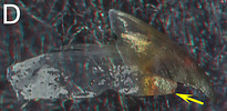
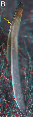
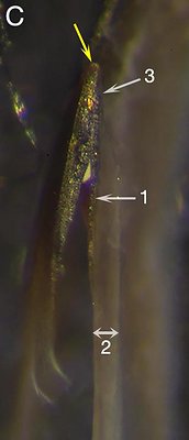
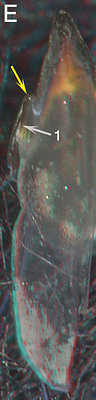
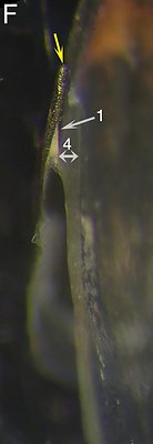
Figure. Lampadioteuthis megaleia. A-C - Female, 32 mm ML, 0.9 mm URL. D-F - Mature male, 25 mm ML, 0.7 mm URL. A & D - Side views showing where the cut was made. B & E - Oral views of entire beak fragment to aid in orientation. C & F - Photomicrograph (2D) of the cut shoulder, oral views taken perpendicular to the plane of the cut.
ARROWS: 1 - Inner Component of Shoulder Blade. 2 - Hyaline material of the Lateral Wall. 3 - Indentation in Shoulder Blade which seems to correspond to thickness of the Inner Component. 4 - Hyaline Matrix. Note the greater thickness compared to the more posterior Lateral Wall. Yellow arrows: Point to the same spot on each beak.In the female L. megaleia (C) the Shoulder Blade consists of two Components. The Inner Component (C, 1) extends posteriorly onto the Lateral Wall and shortly diminishes. The anterior end of this Component is, apparently, marked by small step (C, 3) in the Shoulder Blade. A thick layer of Hyaline Matrix, medial to the Shoulder Blade, sharply tapers toward the anterior tip of the Shoulder Blade (C) and posteriorly is continuous with that of the Lateral Wall (C, 2). In the smaller, mature L. megaleia the Inner Component of the Shoulder Blade (F, 1) is shorter and does not reach the posterior end of the Hyaline Matrix.
No layer of hyaline material is present lateral to the Outer Component of the Shoulder Blade in either beak (C, F). The beaks in both squid have very little Lateral Wall pigmentation and this is very light. No indication of a Palatoshoulder Ridge is present on either beak nor is there a line between the two Components of the Shoulder Blade.
COMMENTS ON 3° UNIPARTITE BEAKS
Three of the five taxa placed in this group (i.e., Slosarczykovia, Joubiniteuthidae, Cranchiinae) are not closely related and their beaks are only similar enough to place them in this "catch-basket" group. We can assume therefore that this similarity is convergent. The remaining two taxa (i.e., Pyroteuthidae, Lampadioteuthinae) are more difficult because (1) they are very small and (2) they may be closely related. If their ancestors were as small as their smallest present members, their size may have posed an evolutionary bottle neck in beak structure resulting in the loss of Tripartite structure. Regarding the second possibility, the structure of the beaks of these two taxa, while showing some similarities, but also some dissimilarities, are not well enough understood to provide additional evidence of a close phylogenetic relationship.


