Back to Profile Page

Tree of Life Media Contributed By David G. Mann

<< < 1 2 3 4 5 6 7 > >>
ID Thumbnail Media Data
29813
Specimen Condition Diagrammatic section of cell wall
Life Cycle Stage Vegetative phase
Body Part Frustule, valve age highlighted
View transverse section
Image Use creative commons This media file is licensed under the Creative Commons Attribution-NonCommercial License - Version 3.0.
Copyright © 2008 David G. Mann
Attached to Group Diatoms: view page image collection
Title cell_division3.jpg
Image Type Diagram
Image Content Body Parts
ID 29813
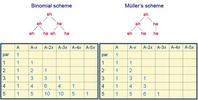
29818 diatom division patterns
Comments Division schemes for diatoms
Life Cycle Stage Vegetative phase
Copyright © 2008 David G. Mann
Image Use restricted
Attached to Group Diatoms: view page image collection
Title division_schemes.jpg
Image Type Diagram
ALT Text diatom division patterns
ID 29818
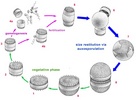
29857 diatom life cycle
Comments diagram of the generalized life-cycle of a centric diatom
Acknowledgements modified from original drawings prepared for F.E. Round et al. (1990: The diatoms. Biology and morphology of the genera. Cambridge University Press, Cambridge.
Life Cycle Stage All
Copyright © 2008 David G. Mann
Image Use restricted
Attached to Group Diatoms: view page image collection
Title diatom_life_cycle_tol.jpg
Image Type Diagram
ALT Text diatom life cycle
ID 29857
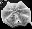
29858 Astermophalus valve
Scientific Name Asteromphalus
Creator Frank E. Round
Acknowledgements This image is derived from the Professor Frank Round Image Archive at the Royal Botanic Garden Edinburgh
Specimen Condition Dead Specimen
Identified By Frank E. Round
Life Cycle Stage Vegetative phase
Body Part valve
View external, SEM
Image Use creative commons This media file is licensed under the Creative Commons Attribution-NonCommercial License - Version 3.0.
Copyright © 2008 David G. Mann
Attached to Group Diatoms: view page image collection
Title asteromphalus814.jpg
Image Type Photograph
Image Content Body Parts, Ultrastructure
ALT Text Astermophalus valve
ID 29858
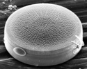
29859 Actinocyclus theca
Scientific Name Actinocyclus
Specimen Condition Dead Specimen
Identified By David G. Mann
Life Cycle Stage Vegetative phase
Body Part theca
View external, SEM
Copyright © 2008 David G. Mann
Image Use restricted
Attached to Group Diatoms: view page image collection
Title actinocyclus.jpg
Image Type Photograph
Image Content Body Parts, Ultrastructure
ALT Text Actinocyclus theca
ID 29859
29860 Arachnoidiscus detail
Scientific Name Arachnoidiscus
Creator Frank E. Round
Acknowledgements This image is derived from the Professor Frank Round Image Archive at the Royal Botanic Garden Edinburgh
Specimen Condition Dead Specimen
Identified By Frank E. Round
Life Cycle Stage Vegetative phase
Body Part valve
View centre internally, SEM
Image Use creative commons This media file is licensed under the Creative Commons Attribution-NonCommercial License - Version 3.0.
Copyright © 2008 David G. Mann
Attached to Group Diatoms: view page image collection
Title arachnoidiscus_ins_centre.jpg
Image Type Photograph
Image Content Body Parts, Ultrastructure
ALT Text Arachnoidiscus detail
ID 29860
29861 Living Coscinodiscus
Scientific Name Coscinodiscus
Identified By David G. Mann
Life Cycle Stage Vegetative phase
Image Use creative commons This media file is licensed under the Creative Commons Attribution-NonCommercial License - Version 3.0.
Copyright © 2008 David G. Mann
Attached to Group Diatoms: view page image collection
Title coscinodiscus_living.jpg
Image Type Photograph
Image Content Specimen(s)
ALT Text Living Coscinodiscus
ID 29861
29862 Rhizosolenia cell
Scientific Name Rhizosolenia
Reference G. Karsten (1905). Das Phytoplankton des Antarktischen Meeres nach dem Material der deutschen Tiefsee-Expedition 1898-1899. Wissenschaftliche Ergebnisse der Deutchen Tiefsee-Expedition auf dem Dampfer "Valdivia" 1898-1899, 2 (2): 1-136.
Creator George Karsten
Acknowledgements scanned from G. Karsten (1905), plate 10, fig. 5d.
Specimen Condition Live Specimen
Life Cycle Stage Vegetative phase
View girdle view
Image Use creative commons This media file is licensed under the Creative Commons Attribution-NonCommercial License - Version 3.0.
Copyright © 2008 David G. Mann
Attached to Group Diatoms: view page image collection
Title rhizosolenia_cell.jpg
Image Type Drawing/Painting
Image Content Specimen(s)
Technical Information as printed, specimen measures 99 mm and represented a magnification of ×250
ALT Text Rhizosolenia cell
ID 29862
29866 Corethron cell
Scientific Name Corethron valdiviae
Reference G. Karsten (1905). Das Phytoplankton des Antarktischen Meeres nach dem Material der deutschen Tiefsee-Expedition 1898-1899. Wissenschaftliche Ergebnisse der Deutchen Tiefsee-Expedition auf dem Dampfer "Valdivia" 1898-1899, 2 (2): 1-136.
Creator George Karsten
Acknowledgements scanned from G. Karsten (1905), plate 12, fig. 2.
Specimen Condition Live Specimen
Identified By George Karsten
Life Cycle Stage Vegetative phase
View girdle view
Image Use creative commons This media file is licensed under the Creative Commons Attribution-NonCommercial License - Version 3.0.
Copyright © 2008 David G. Mann
Attached to Group Corethron: view page image collection
Title corethron_cell.jpg
Image Type Drawing/Painting
Image Content Specimen(s)
Technical Information Cell (without spines) 43.5 mm long in original image, at ×125
ALT Text Corethron cell
ID 29866
29867 Sellaphora valve
Scientific Name Sellaphora
Creator Frieda Christie
Acknowledgements This micrograph was taken by Frieda Christie, Royal Botanic Garden Edinburgh
Specimen Condition Dead Specimen
Identified By David G. Mann
Life Cycle Stage Vegetative phase
Body Part valve
View exterior, SEM
Image Use creative commons This media file is licensed under the Creative Commons Attribution-NonCommercial License - Version 3.0.
Copyright © 2008 David G. Mann
Attached to Group raphid diatoms: view page image collection
Title sellaphora_elliptical.jpg
Image Type Photograph
Image Content Body Parts, Ultrastructure
ALT Text Sellaphora valve
ID 29867
Please note: Most images and other media displayed on the Tree of Life web site are protected by copyright, and the ToL cannot act as an agent for their distribution. If you would like to use any of these materials for your own projects, you need to ask the copyright owner(s) for permission. For additional information, please refer to the ToL Copyright Policies.

Contribute to the ToL

ToL Contributions

Ways to Contribute

Use of Contributions

Scientific Content

Building Treehouses

Images/Media

  go to the Tree of Life home page
top