


Atlantidae
Roger R. Seapy
- Atlanta Lesueur, 1817
- Protatlanta souleyeti (Smith 1888)
- Oxygyrus inflatus Benson 1835
Introduction
The Atlantidae are the most species-rich family of heteropods, containing over 60% of all species. They are microscopic (<1 cm shell diameter), and bear a coiled shell into which their bodies can be retracted, after which the shell aperture is closed off by a chitinous operculum attached to the opercular lobe of the foot. The head has a pair of large tentacles anterior to the eyes. A large muscular sucker, located on the posteroventral margin of the swimming fin, is used to hold prey while feeding. Atlantids are found primarily in the epipelagic zone, between the surface and 200 m and mostly in tropical to subtropical waters. Many species undergo vertical migration from daytime depths into shallower waters at night.
Brief Diagnosis
A family of heteropods with:
- Microscopic size (<1 cm shell diameter)
- Body retractable into coiled shell; shell aperture closed off by chitinous operculum
- Swimming fin with large, muscular sucker that is used to hold prey while feeding
Characteristics
- Shell
- Viewed from the right side, the direction of shell coiling is dextral ("right-handed")
- Shell laterally compressed, with the spire projecting to varied degrees from the right side, except in Oxygyrus inflatus with an involute spire
- A flattened keel of varied height extends outward from the shell's circumference
- Tentacles large and of equal size, extending anteriorly from beneath the eyes
Click on an image to view larger version & data in a new window
Figure. Head, proboscis and tentacles in Atlanta peronii. Tentacles project forward from beneath the eyes on either side of the proboscis. © 2005
- Foot
- Anterior portion of foot forms a muscular swimming fin, with a large sucker on the ventro-lateral margin used to hold prey during feeding and an opercular lobe on the posterior margin of the foot that bears a transparent, chitinous operculum
Click on an image to view larger version & data in a new window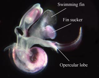
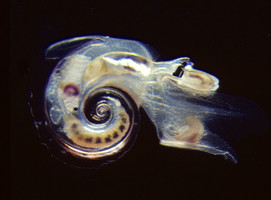
Figure. Oxygyrus inflatus, viewed from the right side. © 2005
- Eye morphology of three types (termed a, b and c; after Richter, 1961). The eye types differ in the pigmented region between the lens and the retina (see labeled drawings below). Type a and b eyes have a dorsal portion of the pigmented wall that is transparent, forming a window. Type c eyes (see also first color photograph below) lack a dorsal window. Type a and b eyes differ in the presence (in the latter) or absence (in the former) of a narrow transverse slit between proximal and distal pigmentation Click on an image to view larger version & data in a new window
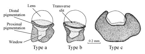
Figure. The three types of eye morphology (types a, b and c) in the Family Atlantidae, drawn in dorsal view. Modified from Richter and Seapy (1999, Fig. 2) © 2005 Roger R. Seapy
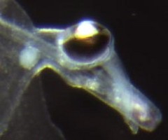
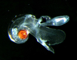
Figure. Type c eye of Atlanta helicinoidea. © 2005
- Operculum
- The operculum is chitinous, thin and flexible, and is attached to the opercular lobe of the foot
- The operculum serves to close off the shell aperture after the animal retracts into its shell
- The operculum consists of an apical gyre (formed during the larval phase) and a distal part that grows downward from the apex (in post-metamorphic animals). When viewed from its inner surface (the side that attaches to the opercular lobe of the foot), the gyre unwinds in a clockwise direction (see sketches below) Click on an image to view larger version & data in a new window
Figure. Sketches of four opercula from Atlanta echinogyra having shell diameters of 0.6, 1.0, 1.2 and 1.5 mm (from left to right). Drawings from Tokioka (1961, fig. 11). © 1961 T. Tokioka
- Three types of opercula, termed a, b and c (see sketches below), were distinguished by Richter (1961) and are found in two (Protatlanta and Atlanta) of the atlantid genera. The type c (monogyre) operculum differs from the type a and b (oligogyre) opercula in the number of turns of the gyre ("single" in the monogyre and "a few" in the oligogyre type). The oligogyre opercula are further distinguished by the relative sizes of their gyres; hence, the prefixes macro- and micro-. The third genus, Oxygyrus, has a markedly different operculum that is broadly triangular and lacks an apical gyre (see the Oxygyrus inflatus page). Click on an image to view larger version & data in a new window
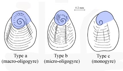
Figure. The three types of opercular morphologies (a, b, c) in the Atlantidae, excepting Oxygyrus. The apical (larval) portion of the operculum is shaded blue. Modified from Richter and Seapy (1999; Fig. 3). © 2005
- The opercular gyre lacks ornamentation in Protatlanta and all species of Atlanta except for A. plana, A. echinogyra and A. turriculata, which possess distinctive gyre spination
- Radula
- Radula with multiple rows of teeth; each row consisting of a central (rachidian) tooth that is flanked on each side by a lateral tooth and two marginal teeth (see SEM image below)
- Radular morphology of two distinctively different types (termed I and II by Richter, 1961). A discussion of the differences between the two types (seen in all heteropods) is given on the Pterotracheoidea page. The radulae of Oxygyrus inflatus, Protatlanta souleyeti are type I; while among the species of Atlanta, eleven (belonging to the A. inflata, A. lesueurii and A. gaudichaudi species groups) are type I and eight (in the A. peronii, A. inclinata and A. gibbosa species groups) are type II (see the Atlanta page)
Click on an image to view larger version & data in a new window
Figure. Scanning electron micrograph of the radula in Atlanta californiensis. © 1993 G. Richter.
- Central (rachidian) tooth with one (Atlanta and Protatlanta) or three (Oxygyrus) pointed cusps
Click on an image to view larger version & data in a new window
Figure. Sketches of the rachidian (central) teeth in the three genera of the family Atlantidae; examples from Atlanta peronii, Protatlanta souleyeti and Oxygyrus inflatus. From Richter (1961, figs. 2a,b,c). © 1961 G. Richter
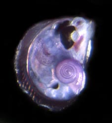

Figure. Left: Atlanta helicinoidea viewed from right side, with body retracted into shell. Right: Scanning electron micrograph of Atlanta plana, with shell tilted to illustrate aperture and spire shape. Shell diameter (right) = 2.1 mm. © 2005
Comments
The family Atlantidae includes three genera, two of which (Protatlanta and Oxygyrus) are monotypic. The genera can be distinguished by the following characters:
| Genus | Shell and keel compositon | Shell spire | Spiral portion of operculum |
|---|---|---|---|
| Atlanta | Calcareous shell and keel | Present (evolute) | Present |
| Protatlanta | Calcareous shell and conchiolin* keel | Present (evolute) | Present |
| Oxygyrus | Conchiolin* shell and keel | Absent (involute) | Absent |
* Conchiolin, especially in fresh specimens, has a glass-like transparency (see title illustration)
Because of the presence of a shell, sinking should be a major problem in the atlantids. As noted elsewhere, the carinariids and pterotracheids have enlarged, elongated bodies containing gelatinous tissues in which heavier sulfate ions are replaced by lighter chloride ions to achieve neutral buoyancy. Laboratory observations of Oxygyrus inflatus by Land (1982) have shown that animals will alternately swim upwards for several seconds and then sink back down with their bodies extended from their shells for about 10 seconds. In-situ field observations of atlantids at night by Gilmer (in Lalli and Gilmer, 1989) indicated very different behavior ftom that exhibited during the day; individuals were motionless, attached to long strands of mucus that appear to originate from the foot.
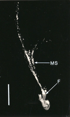
Figure. In-situ photograph of an unidentified atlantid attached to mucus strands (= MS), that extend about 45 mm above the animal. Note that the mucus appears to come from the foot (= F). Photograph modified from Lalli and Gilmer (1989, fig. 11). Scale bar = 10 mm. © 1989 Ronald Gilmer
References
Lalli, C. M. and R. W. Gilmer. 1989. Pelagic snails. The biology of holoplanktonic gastropod snails. Stanford University Press, Stanford. 259 pp.
Land, M. F. 1982. Scanning eye movements in a heteropod mollusc. Journal of Experimental Biology 96: 427-430.
Richter, G. 1961. Die Radula der Atlantiden (Heteropoda, Prosobranchia) und ihre Bedeutung für die Systematik und Evolution der Familie. Zeitschrift für Morphologie und Ökologie der Tiere 50: 163-238.
Richter, G. and R. R. Seapy. 1999. Heteropoda, pp. 621-647. In: D. Boltovskoy (ed.), South Atlantic Zooplankton. Backhuys Publishers, Leiden.
Seapy, R. R. 1990. The pelagic family Atlantidae (Gastropoda: Heteropoda) from Hawaiian waters: a taxonomic survey. Malacologia 32: 107-130.
Spoel, S. van der. 1976. Pseudothecosomata, Gymnosomata and Heteropoda (Gastropoda). Bohn, Scheltema and Holkema, Utrecht. 484 pp.
Spoel, S. van der, L. Newman and K. W. Estep. 1997. Pelagic molluscs of the world. World Biodiversity Database, CD-ROM Series. Expert Center for Taxonomic Identification (ETI), University of Amsterdam. UNESCO Publishing, Paris.
Title Illustrations

| Location | Hawaiian waters |
|---|---|
| Specimen Condition | Live Specimen |
| Sex | Female |
| Life Cycle Stage | adult |
| View | right side |
| Image Use |
 This media file is licensed under the Creative Commons Attribution-NonCommercial License - Version 3.0. This media file is licensed under the Creative Commons Attribution-NonCommercial License - Version 3.0.
|
| Copyright |
©

|

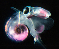
| Scientific Name | Protatlanta souleyeti |
|---|---|
| Location | Hawaiian waters |
| Specimen Condition | Live Specimen |
| Life Cycle Stage | adult |
| View | right side |
| Image Use |
This media file is licensed under the Creative Commons Attribution-NonCommercial License - Version 3.0.
|
| Copyright |
©
|

| Location | Hawaiian waters |
|---|---|
| Specimen Condition | Live Specimen |
| Sex | Female |
| Life Cycle Stage | adult |
| View | right side |
| Image Use |
This media file is licensed under the Creative Commons Attribution-NonCommercial License - Version 3.0.
|
| Copyright |
©
|
About This Page
California State University, Fullerton, California, USA
Correspondence regarding this page should be directed to Roger R. Seapy at
Page copyright © 2010
 Page: Tree of Life
Atlantidae .
Authored by
Roger R. Seapy.
The TEXT of this page is licensed under the
Creative Commons Attribution License - Version 3.0. Note that images and other media
featured on this page are each governed by their own license, and they may or may not be available
for reuse. Click on an image or a media link to access the media data window, which provides the
relevant licensing information. For the general terms and conditions of ToL material reuse and
redistribution, please see the Tree of Life Copyright
Policies.
Page: Tree of Life
Atlantidae .
Authored by
Roger R. Seapy.
The TEXT of this page is licensed under the
Creative Commons Attribution License - Version 3.0. Note that images and other media
featured on this page are each governed by their own license, and they may or may not be available
for reuse. Click on an image or a media link to access the media data window, which provides the
relevant licensing information. For the general terms and conditions of ToL material reuse and
redistribution, please see the Tree of Life Copyright
Policies.
- First online 16 February 2005
- Content changed 01 April 2010
Citing this page:
Seapy, Roger R. 2010. Atlantidae . Version 01 April 2010 (under construction). http://tolweb.org/Atlantidae/28732/2010.04.01 in The Tree of Life Web Project, http://tolweb.org/


