


Chiroteuthis picteti
Clyde F. E. Roper and Richard E. Young

Introduction
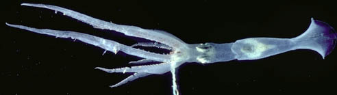
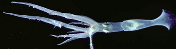
Chiroteuthis picteti is a medium-sized deep-sea squid with a broad distribution in the Pacific and Indian Oceans. It is separated most easily from most other members of the genus by its very long, slender, tentacular clubs, a feature shared with C. mega.
Brief diagnosis:
A Chiroteuthis ...
- with three rows of individual, round photophores on each eyeball.
- with proximal region of club very short.
Characteristics
- Arms
- Large suckers with 10-20 usually separate, pointed to blunt teeth on distal 2/3 of ring.
- Largest suckers not globular.
- Tentacular clubs
- Suckers with central tooth enlarged.
- Sucker stalks divided into two parts with flag-like lateral keel on proximal segment; stalks of suckers in lateral series about twice as long as those of medial series.
- Protective membranes
- Protective membranes divide club into two portions with proximal portion set less than one tenth length of distal portion and with proximal trebeculae only slightly longer than distal trabeculae.
Click on an image to view larger version & data in a new window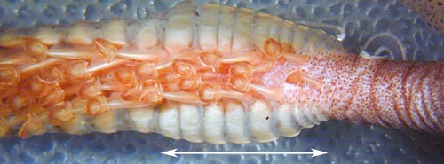
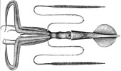
Figure. Oral view of club base of C. picteti showing short proximal section of the protective membrane (indicated by arrow). Photograph by R. Young.
- Protective membranes divide club into two portions with proximal portion set less than one tenth length of distal portion and with proximal trebeculae only slightly longer than distal trabeculae.
- Head
- Beaks: Descriptions can be found here: Lower beak; upper beak.
- Beaks: Descriptions can be found here: Lower beak; upper beak.
- Photophores
- Eyeball- 3 series of round photophores: lateral series= 6 to 9; intermediate series = 8 to 11; medial series = 6 to 10; terminal photophores slightly larger.
- Viscera: two photophores.Click on an image to view larger version & data in a new window
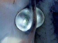
Figure. C. picteti, female, nearly mature. ? mm ML. Ventrolateral view of visceral photophores (fresh ?). Photograph by R. Young and C. Roper.
- Pigmentation
- Club sucker stalks without pigment.
- Buccal membrane usually with dense epidermal pigment.
Comments
More details of the description of C. picteti can be found here.Nomenclature
C. picteti was originally described from Indonesian waters by Joubin (1894). Shortly thereafter Goodrich (1896) described two species from the Bay of Bengal, Indian Ocean. One, based on a large adult (360 mm ML) was named C. macrosoma. The other, based on a doratopsis paralarva, was named C. pellucida. Both of these species appear to be synonyms of C. picteti. Chun (1908) described C. imperator from off Sumatra, Indonesia, close to the type locality of C. picteti. Our examination of the type of C. imperator showed no distinguishing features, and we consider this species to be a synonym of C. picteti. Salcedo-Vargas (1996) described a subspecies, C. picteti somaliensis, from the western Indian Ocean that is distinguished by having more than the typical number of teeth on the arm sucker rings (18-22) and the tentacle-club sucker rings (13-15).
Life history
Paralarval doratopsid stages have been described from Hawaiian waters (as C. imperator) by Young, 1991. The younger stages are unusual for a doratopsis in virtually lacking a brachial pillar and, at later stages, having the esophagus centrally located in the now present brachial pillar. The advanced paralarva reaches a length of at least 45 mm ML.

Figure. Paralarval stages of C. picteti from Hawaiian waters (Young, 1991): A - Side view of head, 2.4 mm ML; note the virtual absence of a brachial pillar at this stage. The esophagus within the brachial pillar (apparent in later stages) occupies a central position. B - Ventral and dorsal views, 2.8 mm ML. C - Ventral and dorsal views, 7.7 mm ML. D - Ventral and dorsal views, 12 mm ML. The median chromatophore between the eyes on the ventral surface of the head is an important systematic character. Scale bar is 1 mm. Drawings from Young (1991).
Spent females are rarely found in the Chiroteuthididae. We describe one observation here. A spent female C picteti, dip-netted by a fisherman off Oahu Hawaii, was seen swimming at the surface in mid-afternoon over bottom depths of 2,000 m (8 mi. offshore). The squid was 370 mm ML and the tissues were quite flaccid. No trace of discharged spermatophores or spermathecae could be found. Virtually no trace of the ovary remained except for a few spherical eggs measuring about 1.0 mm in diameter. The squid was reddish-brown in color with most pigment in chromatophores. One exception, however, was the oral surface of the ventral arms (arms IV) including the oral surface of the tentacular sheath. these surfaces had a very dark pigmentation with much of the pigment in epithelial cells. The pigmentation here was much darker than anywhere else on the squid. (The usually heavily pigmented buccal membrane was not as dark as in immature squid).
A mature male that we examined, captured in a trawl, had no such specialized pigment on the ventral arms. Considering the luminous organs associated with the tentacles, perhaps the pigmentation acts as a shutter over the retracted tentacles that are involved in some luminescent reproductive behavior. The male (200 mm ML), which lacked hectocotylization of the arms, had a large penis that extended well outside the mantle cavity. A few spermatophores had been discharged, apparently during capture. One of these was found attached inside but near the tip of the funnel and suggests that normally the penis is extended through the funnel.
Distribution
We have observed considerable geographical variability in this species. More careful study could indicate that this is a species complex. At present, however, the data warrant recognition of only a single species. This species is found in the tropical and subtropical Indo-West Pacific.
References
Chun, C. 1908. Ueber Cephalopoden der Deutschen Tiefsee-Expedition, Zoologischer Anzeiger, 33: 86-89.
Goodrich E. S. 1896. Report on a collection of Cephalopoda from the Calcutta Museum. London, Transactions of the Linnean Society, series 2, 7: 1-24.
Joubin, L. 1894. Note p;reliminaire sur les Cephalopodes provenents des campagnes du Yacht, L’Hirondelle. Memoires de la Societe Zoologique de France, 7: 211-216.
Salcedo-Vargas, M. A. 1996. Cephalopods from the Netherlands Indian Ocean Programme (NIOP) - I. Chiroteuthis spoeli, n. spec. and Chiroteuthis picteti somaliensis n. subspec. Beaufortia, 46: 11-26.
Young, R. E. 1991. Chiroteuthid and related paralarvae from Hawaiian waters. Bull. Mar. Sci., 49: 162-185.
Title Illustrations
| Scientific Name | Chiroteuthis picteti |
|---|---|
| Location | Hawaii |
| Life Cycle Stage | young subadult |
| Scientific Name | Chiroteuthis picteti |
|---|---|
| Location | Eltanin??? |
| Creator | J. Schroeder |
About This Page

Smithsonian Institution, Washington, D. C., USA
University of Hawaii, Honolulu, HI, USA
Page copyright © 2019 and
All Rights Reserved.
- Content changed 10 October 2017
Citing this page:
Roper, Clyde F. E. and Richard E. Young. 2017. Chiroteuthis picteti . Version 10 October 2017 (under construction). http://tolweb.org/Chiroteuthis_picteti/19477/2017.10.10 in The Tree of Life Web Project, http://tolweb.org/


