


Histioteuthis atlantica
Richard E. Young and Michael Vecchione

Introduction
H. atlantica has a circumglobal distribution in southern waters and reaches a size of, at least, 258 mm ML.
Characteristics
- Photophores
- Large, compound photophores with anteriorly and posteriorly (see black arrows in photograph to the right) directed reflectors .
- Arms IV with 4 longitudinal series on arm base (dorsal series with organs of mixed sizes).
- Terminal third of each arm I-III with row of large, elliptical, darkly pigmented, simple organs.
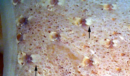
Figure. Ventral view of an anterior segment of the ventral mantle of H. atlantica, 57 mm ML, 48°S, 145°E, USNM cat. no. 814656. Black arrows point to the Posterior reflector of large photophores; white arrows point to small photophores. Photograph by R. Young.
Comments
More details of the description can be found here.Species of the reversa-group are distinguished by the following characters:
- Compound photophores
- Large and small photophores intermixed on ventral surface of mantle .
- 18 photophores (17 large and 1 small) around right eyelid.
- Beak
- Weakly developed median ridge on each lateral wall.
- Tubercles
- Absent.
This species is most easily separated from other members of the reversa group by (1) the arrangement of photophores on arms IV (H. reversa - 4 series, dorsal one with all small organs; H. eltaninae - 3 series), (2) the absence of specialized arm-tip photophores in H. eltaninae and H. reversa.
The posteriorly directed reflectors are also known in H. reversa, but the condition in H. eltaninae is unknown. With the exception of information on the posterior reflectors of the compound photophores, the above information is from Voss (1969).
Life History
Numerous small, darkly pigmented, simple photophores on the posterior region of the mantle only appear at maturity. The number and density of the photophores increases with size of the squid. Mature females are unknown. (Voss, et al., 1998)

Figure. Ventral and dorsal views of the mantle of a mature male of H. atlantica, 115 mm ML, 40° 18'S, 39° 12'W. Drawing from Voss, et al., 1998..
Distribution
Geographical distribution
Histioteuthis atlantica exhibits a circumglobal southern distribution. Its distribution strongly overlaps with the more southern distribution of its close relative H. eltaninae. Histioteuthis atlantica is regularly found over ocean basins, shelf and plateau areas. (Voss, et al., 1998)

Figure. Chart or the geographical distribution of H. atlantica. Modified from Voss, et al., 1998.
References
Voss, N. A. 1969. A monograph of the Cephalopoda of the North Atlantic: The family Histioteuthidae. Bull. Mar. Sci., 19: 713-867.
Voss, N.A., K. N. Nesis, P. G. Rodhouse. 1998. The cephalopod family Histioteuthidae (Oegopsida): Systematics, biology, and biogeography. Smithson. Contr. Zool., 586(2): 293-372.
Title Illustrations

| Scientific Name | Histioteuthis atlantica |
|---|---|
| Location | 43° 45''S, 174° 07''W |
| Reference | from Voss, N. A. 1969. A monograph of the Cephalopoda of the North Atlantic: The family Histioteuthidae. Bull. Mar. Sci. 19:713-867. printed with permission. |
| Sex | Male |
| View | ventral |
| Size | 53 mm ML |
| Copyright | © 1969 Bulletin of Marine Science |

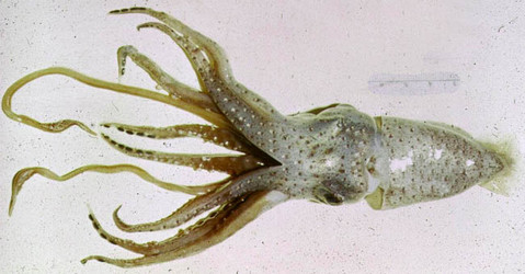
| Scientific Name | Histioteuthis atlantica |
|---|---|
| Comments | Most of the chromatophores in the skin of this squid were contracted when the photograph was taken. This squid appears silvery, a condition that is unusual in histioteuthids. |
| Specimen Condition | Fresh |
| View | Ventral |
| Copyright | © E. S. McSweeny |
About This Page

University of Hawaii, Honolulu, HI, USA
National Museum of Natural History, Washington, D. C. , USA
Page copyright © 2013 and
 Page: Tree of Life
Histioteuthis atlantica .
Authored by
Richard E. Young and Michael Vecchione.
The TEXT of this page is licensed under the
Creative Commons Attribution-NonCommercial License - Version 3.0. Note that images and other media
featured on this page are each governed by their own license, and they may or may not be available
for reuse. Click on an image or a media link to access the media data window, which provides the
relevant licensing information. For the general terms and conditions of ToL material reuse and
redistribution, please see the Tree of Life Copyright
Policies.
Page: Tree of Life
Histioteuthis atlantica .
Authored by
Richard E. Young and Michael Vecchione.
The TEXT of this page is licensed under the
Creative Commons Attribution-NonCommercial License - Version 3.0. Note that images and other media
featured on this page are each governed by their own license, and they may or may not be available
for reuse. Click on an image or a media link to access the media data window, which provides the
relevant licensing information. For the general terms and conditions of ToL material reuse and
redistribution, please see the Tree of Life Copyright
Policies.
- Content changed 03 November 2013
Citing this page:
Young, Richard E. and Michael Vecchione. 2013. Histioteuthis atlantica . Version 03 November 2013 (under construction). http://tolweb.org/Histioteuthis_atlantica/19789/2013.11.03 in The Tree of Life Web Project, http://tolweb.org/


