Onychoteuthis
Michael Vecchione, Richard E. Young, and Kotaro Tsuchiya


This tree diagram shows the relationships between several groups of organisms.
The root of the current tree connects the organisms featured in this tree to their containing group and the rest of the Tree of Life. The basal branching point in the tree represents the ancestor of the other groups in the tree. This ancestor diversified over time into several descendent subgroups, which are represented as internal nodes and terminal taxa to the right.

You can click on the root to travel down the Tree of Life all the way to the root of all Life, and you can click on the names of descendent subgroups to travel up the Tree of Life all the way to individual species.
For more information on ToL tree formatting, please see Interpreting the Tree or Classification. To learn more about phylogenetic trees, please visit our Phylogenetic Biology pages.
close boxIntroduction
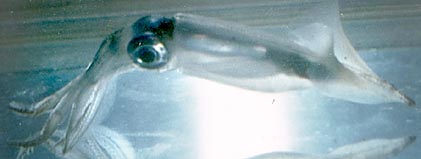
Members of the genus are mostly small to medium sized squids of under 20 cm in mantle length as adults. The largest, however, O. borealijaponicus, reaches a size of 350 mm ML (Kubodera, et al., 1998). Species are found mostly in tropical and subtropical waters throughout the world's oceans although they are also common in high latitudes of the North Pacific where O. borealijaponicus reaches subarctic waters. One of the diagnostic features of Onychoteuthis species is the presence of a bilobed photophore on the ventral surface of each eyeball as seen in the photograph below right. Another diagnostic feature is the presence of two photophores (arrows) on the viscera as can barely be seen through the mantle in the photograph below left.
 image info
  image info
 Figure. Ventral views of Onychoteuthis sp. showing photophores. Left - Arrows indicate visceral photophores (ocular photophores also visible), juvenile. Right - Ocular photophores of a different squid. Both squid from Hawaii waters. Photographs by R. Young.
Species of Onychoteuthis are commonly seen in surface waters at night and are often collected by dipnet at nightlight stations. Only young squid are normally captured in standard midwater trawls; apparently older squids avoid the trawls. One non-standard method of collection results from individuals being able to leap high out of the water and, sometimes, landing on the deck of a ship.
Diagnosis
An onychoteuthid ...
- with photophores (unique character).
- with gladius visible beneath skin in dorsal midline.
- with 8-13 occipital folds.
Characteristics
- Arms
- Distal, fleshy thickening on each sucker (see arrows in photograph below). This delicate thickening or knob is not obvious on all suckers and when present it is easily damaged during capture.
- Protective membranes low and with trabeculae fused to sucker base and not clearly seen as separate entities (see photograph below).
 image info
  image info
Figure. xxLateral views of preserved arm suckers of Onychoteuthis sp. Photographs by R. Young.
- Tentacular club
- Marginal suckers absent in subadults except for O. meridiopacifica (several marginal suckers present).
- Dorsal series of hooks with hooks that are small in size in mid-series (see arrows on drawing below) with larger hooks proximally and distally.
 image info
Figure. Oral view of tentacular club of Onychoteuthis "banksi." Drawing modified from Naef, 1921/23.
- Head
- 8-13 occipital folds present on each side of head. Arrows in the photograph below indicate the ventral 7 occipital folds. Additional very small dorsal folds are out of focus and not visible in the picture. The olfactory organ is at the posterodorsal end of occipital fold II.
 image info
Figure. Lateral view of head showing occipital folds of preserved Onychoteuthis sp. Photograph by R. Young.
- 8-13 occipital folds present on each side of head. Arrows in the photograph below indicate the ventral 7 occipital folds. Additional very small dorsal folds are out of focus and not visible in the picture. The olfactory organ is at the posterodorsal end of occipital fold II.
- Funnel groove
- Funnel groove with distinct ridges along margin; margins convex near anterior, pointed end of groove (squid must be in good condition to see this feature).
 image info
Figure. Ventral view showing funnel groove of preserved Onychoteuthis sp. Photograph by R. Young.
- Funnel groove with distinct ridges along margin; margins convex near anterior, pointed end of groove (squid must be in good condition to see this feature).
- Photophores
- Large organ present ventrally on each eye (see Introduction).
- Two organs present on intestine.
 image info
Figure. Ventral view of intestinal photophores of Onychoteuthis sp. Drawing modified from Young, 1972.
- Gladius
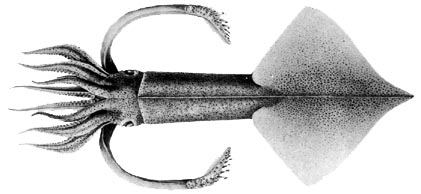
- The dorsal mantle musculature and fins are bisected by the gladius. (The resulting line down dorsal midline generally is visible through the skin - see title drawing).
- Rostrum thin (i.e., laterally compressed) and pointed (except in O. meridiopacifica).
Species comparisons
| Species | Posterior visceral photophore | Number of club hooks | Marginal club suckers | Maximum size | Habitat |
| O. banksii | Large, round | 22 | None | ? | Off tropical W. Africa |
| O. borealijaponicus | Large, slender | 24-27 | None | 350 mm ML | High N. Pacific |
| O. compacta | Large, round | ??? | None | ? | Hawaiian waters |
| O. meridopacifica | Small, round | 16-19 | Few | 70 mm ML | Tropical S. Pacific |
Behavior
Paralarvae of Onychoteuthis, like the paralarvae of many other squid, have the ability to withdraw their head and arms into their mantle cavity. This behavior is, presumably, defensive.
Figure. Ventral views of a paralarva of O. compacta, head extended and retracted, off Hawaii. Photographs.
Life History
Most paralarval Onychoteuthis are easily recognized by the pointed rostrum on the gladius. If the fins are damaged, the rostrum projects well beyond the fins (see photographs of the paralarvae in the previous section). The photograph on the left (ventral view, posterior end of a paralarva) shows the rostrum barely emerging and the photograph on the right (side view, posterior end of a paralarva) shows the rostrum just reaching to the edge of the fins. Even if the rostrum is not protruding due to damage, it still can be easily seen.
 image info
  image info
Figure. Ventral and side views of posterior ends of Onychoteuthis sp. paralarva, off Hawaii. Photographs by R. Young.
The adult female looses its tentacles apparently near the time of spawning. Spent females which are very flaccid were originally described as belonging to the genus Chaunoteuthis. In the photograph below the upper white arrow points to a scar that parallels much of the sissors cut through the mantle. This scar was apparently made by the male when mating. The lower arrow points to one of about 9 spermatangia embedded in the mantle muscle that apparently entered at the scar.
 image info
  image info
Figure. Left - Ventral view, female "chunoteuthis stage" of Onychoteuthis sp. , a mated and presumably (i.e. damage prevented assessment) spent female, preserved, 155 mm ML. Photograph by R. Young. Right - Dorsal view of "Chunoteuthis mollis". Drawing from Pfeffer (1912).
Distribution
Species of Onychoteuthis are absent from the Arctic, upper boreal Atlantic, subantarctic and Antarctic waters, but occur in all other oceans (Nesis, 1982/87).
References
Kubodera, T., U. Piakowski, T. Okutani and M. R. Clarke. 1998 Taxonomy and zoogeography of the family Onychoteuthidae. Smiths. Contr. to Zoology, No. 585: 277-291.
Nesis, K. N. 1982/87. Abridged key to the cephalopod mollusks of the world's ocean. 385+ii pp. Light and Food Industry Publishing House, Moscow. (In Russian.). Translated into English by B. S. Levitov, ed. by L. A. Burgess (1987), Cephalopods of the world. T. F. H. Publications, Neptune City, NJ, 351pp.
Young, R. E. 1972. The systematics and areal distribution of pelagic cephalopods from the seas off Southern California. Smithson. Contr. Zool., 97: 1-159.
About This Page
National Marine Fisheries Service
Systematics Laboratory
National Museum of Natural History
Washington, D. C. 20560
USA
Richard E. Young
Dept of Oceanography
University of Hawaii
Honolulu, Hawaii 96822
USA
Tokyo University of Fisheries, Konan, Minato, Tokyo
Page copyright © 2003 , Richard E. Young, and
Citing this page:
Vecchione, Michael, Young, Richard E., and Tsuchiya, Kotaro. 2003. Onychoteuthis . Version 01 January 2003 (under construction). http://tolweb.org/Onychoteuthis/19955/2003.01.01 in The Tree of Life Web Project, http://tolweb.org/


