- Photophores (head)
- Compound photophores (Type 1b):
 Click on an image to view larger version & data in a new window
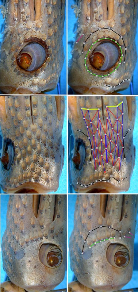
Figure. Various views of the head photophores of H. celetaria. Right photographs - Show one or more of the basic photophore groups, from different views, with superimposed lines and dots to emphasize the photophore groups. Left photographs - Show the same picture without superimposed lines and dots so that the photophores can be seen without obstruction. The superimposed lines are formed by first placing white dots over the pigmented, photogenic cups of all photophores in a particular group. The dots are then joined by a line of the appropriate color. Photographs by R. Young.
- Basal Arm IV Row with 3 photophores. Medial photophore separated by 1 photophore from anterior photophore of Midline Series.
- Midline Series with 3 photophores.
- Right Eyelid Series with 17 photophores (rarely 19 or 18: Voss, et al., 1998).
- Left Eyelid Series with 6 photophores.
- 2° Right Eyelid Series with 8 photophores.
- 2° Left Eyelid Series with 7 photophores.
- Ventral Matrix with 36 photophores including an asymmetrically placed dangling, rogue photophore (colored red in illustration), and 9 longitudinal (blue) series. Attachment to 2° Eyelid Series at 2° eyelid photophores no. 2 either side.
- Ventral Matrix photophores do not align in transverse rows.
- Right basal series absent.
- Left basal series apparently with two small photophores.
- Basal row with 9 photophores and one sawtooth (note that two of the sawtooth photophores align with the rogue (red) photophore..
- Right Accessory Series with 2 photophores (see below).
- Left Accessory Series with 3 photophores (see below).
Click on an image to view larger version & data in a new window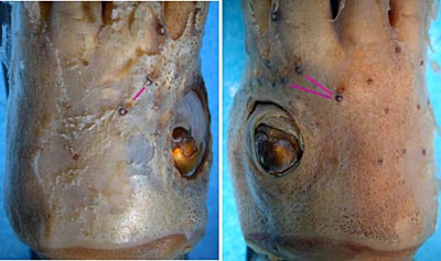
Figure. Dorsal view of the head photophores of H. celetaria. Photographs by R. Young.
- Simple photophores (round, black dots):
- Arrangement as illustrated.
- Large, simple photophores on dorsal surface of head absent.
- Small, simple photophores not easy to recognize and may be variable.
- Compound photophores (Type 1b):
- Mantle photophores.
- Compound photophores of large, uniform size and evenly spaced on anterior 2/3 of ventral mantle.
Click on an image to view larger version & data in a new window
Figure. Ventral view of mantle of H. celetaria, holotype, 39 mm ML. Extracted from Fig. 15a of Voss (1969).
- Arm photophores.
- Arms IV with 3 longitudinal series of compound photophores on arm base.
- Group of 4-8 compound photophores on ends of all arms in immatures but only on arms IV in mature squid.
- Long, narrow, black, simple photophore beneath keel on distal portions of each arm I-III in mature squid. The drawing to the right shows a cross-section through the aboral region of arm I.
Click on an image to view larger version & data in a new window
Figure. Ventral view of arm IV of H. celetaria, holotype. Drawing from Voss, 1969 (Fig 15f).
Click on an image to view larger version & data in a new window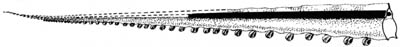
Figure. Lateral view of arm I (reconstructed) of H. celetaria showing simple, aboral photophore that was sectioned in the adjacent drawing. Drawing from Voss, et al., 1998.
Click on an image to view larger version & data in a new window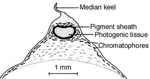
Figure. Cross-sectional view of the aboral photophore of H. celetaria, 258 mm ML mature female, 13° 07'S, 9° 02'W. Drawing from Voss, et al., 1998.
- Tentacles
- Suckers on manus closely arranged; median 2-3 rows only slightly enlarged.
Click on an image to view larger version & data in a new window
Figure. Oral views of the tentacular club of H. celetaria. Top - Holotype, 39 mm, female from 32° N,65° W. Drawing from Voss, 1969 (Fig. 15e). Bottom - 74 mm female from 19° S, 4° W. Drawing from Voss, et al. (1998).
- Arms
- Approximately equal to mantle in length.
- Approximately equal to mantle in length.
- Sucker dentition
- Largest suckers of manus with 12-13 sharp teeth on distal margin, proximal margin smooth (drawing to the near right).
- Suckers of 2-3 ventral-marginal series with asymmetrical outer rings giving the sucker a triangular appearance (drawing to the far right). This asymmetry is most pronounced in subadults.
- Large arm suckers with smooth rings except for an occasional sucker on arms IV.
Click on an image to view larger version & data in a new window
Figure. Left - Oral view of largest club sucker of H. celetaria, holotype. Drawing from Voss, 1969 (Fig 15c). Right - Oral view of club sucker,ventral series, same club. Drawing from Voss, 1969 (Fig 15b).
- Occipital folds
- Two prominent occipital folds present on each side of head; ventral fold sharply curved dorsally, terminating in olfactory organ.
Click on an image to view larger version & data in a new window
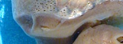
Figure. Lateral view of occipital fold of H. celetaria. Left - Holotype. Drawing from Voss, 1969 (Fig 15g). Right - NMNH specimen. Photograph by R. Young.
- Funnel organ
- Dorsal pad of funnel organ unsculptured.
Click on an image to view larger version & data in a new window
Figure. Ventral view of funnel organ of H. celetaria, holotype. Drawing from Voss, 1969 (Fig. 15h).
- Gladius
- Anterior shoulders of vanes flare and angular.
Click on an image to view larger version & data in a new window
Figure. Ventral view of gladius of H. celetaria, 87 mm GL, mature male, 18° 36'S, 4° 18'W. Drawing from Voss, et al., 1998.
- Fins
- Length about 50-55% of ML; width about 68-72% of ML.
- Length about 50-55% of ML; width about 68-72% of ML.
- Spermatophores
- Not known.
- Not known.
- Hectocotylus
- Ends of arms I modified with equal-sized suckers on long pedicels. Large mature males with suckers on basal portions of all arms enlarged and with swollen, fleshy collars.
Comments
Other than the head-photophore information and the photographs, this description is from Voss (1969) and Voss, et al. (1998).


