


Mastigoteuthidae
Mastigoteuthis
Whip-lash squid
Michael Vecchione, Richard E. Young, and Mario Alejandro Salcedo-Vargas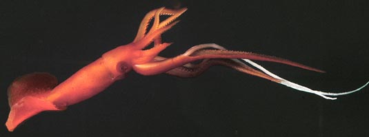


This tree diagram shows the relationships between several groups of organisms.
The root of the current tree connects the organisms featured in this tree to their containing group and the rest of the Tree of Life. The basal branching point in the tree represents the ancestor of the other groups in the tree. This ancestor diversified over time into several descendent subgroups, which are represented as internal nodes and terminal taxa to the right.

You can click on the root to travel down the Tree of Life all the way to the root of all Life, and you can click on the names of descendent subgroups to travel up the Tree of Life all the way to individual species.
For more information on ToL tree formatting, please see Interpreting the Tree or Classification. To learn more about phylogenetic trees, please visit our Phylogenetic Biology pages.
close boxIntroduction
Mastigoteuthids are deep water pelagic or benthopelagic squids that are morphologically distinctive. They are weakly muscled and reddish in color and have elongate fourth arms. Much of the red pigment is not in chromatophore organs but dispersed in other integumental cells, although chromatophores are present. The Mastigoteuthidae, however, is among the most taxonomically confused families of all deep-sea squid. The reason for the taxonomic confusion within the family is that many characters are based on the structure of the tentacles and photophores in the skin. Tentacles are often lost and the skin abraded during capture.
The tentacles are elongate and whip-like with tentacular clubs that are little differentiated from the tentacular stalks except that they are covered with thousands of extremely small suckers; depending on the species, the suckers may be invisible to the naked eye. The photograph below shows the midregion of a tentacular club with the sucker-bearing portion marked by an X. The arrow points to a microscope enlargment showing the small suckers. The tentacles appear, in freshly dead specimens, to function much like "fly paper": anything that touches them sticks.
 image info
 Figure. Side view of a portion of the tentacle club (bottom) of Mastigoteuthis sp., Hawaiian waters, showing the club covering the top half of the cyclindrical tentacle. Insert - Portion of the club, marked by an "X", as seen through a microscope. Photographs by R. Young.
Histological sections through the tentacle can be seen here.
Fins are usually very large and positioned mostly posterior to the muscular part of the mantle. No well-developed system of giant nerve fibers is present, which reflects the absence of rapid jet propulsion (Dilly, et al., 1977).
Diagnosis
Member of the chiroteuthid families ...- with arms IV longest.
- with cyclindrical, whip-like tentacular clubs bearing small suckers in numerous irregular series.
- with oval funnel locking-appartus usually bearing various knobs or lobes (often a tragus and antitragus) affecting the shape of the groove.
Characteristics
- Arms
- Arms IV longest, thickest and with expanded lateral membranes which form tentacular sheaths.
- Tentacles
- Tentacular clubs elongate, virtually cyclindrical; with suckers in many irregular series (30 or more in some species).
- Funnel
- Funnel locking-apparatus oval, usually with knobs (tragus, antitragus) affecting the shape of the depression in the funnel component in different species; specific shape varies with species.
- Fins
- Fins large (ca. 50% of ML) to very large (ca. 90% of ML).
- Fins with terminal position.
- Tail
- Short tail present (often absent due to damage during capture).
- Tail supported by secondary conus.
- Photophores
- Photophores present on eyeball or eyelid and/or integument or absent.
Comments
Integumental photophores are found in many species and seem have a similar structure. The photophore has a large covering chromatophore, a thick donut-shaped cup of photocytes and a central cord of cells that passes through the hole in the photocyte donut to the chromatophore.
 image info
 Figure. Composite longitudinal section through an integumental photophore of M. flammea. Drawing modified from Chun (1910).
The integument of most species (M. pyrodes is an exception) with integumental photophores is complex and contains two elements in addition to the integumental photophores: (1) very small, whitish balls and (2) whitish rings surrounding chromatophores. Since the function of these two structures is unknown, we call the first "white balls" and the second "ringed chromatophores" for descriptive purposes.
 image info
 Figure. Ventral view of the head integument of M. flammea, Gulf of Mexico. Note that much of the pigmentation does not lie within chromatophores. Bottom arrow - A typical integumental photophore. Middle arrow - A very small, light colored ball (a "white ball"). Top arrow - A chromatophore surrounded by a light-colored band (a "ringed chromatophore"). The band is not a shrinkage space around the chromatophore nor is the combination of the band and chromatophore a developmental stage of a typical integumental photophore. Photograph by R. Young.
Comparison of species
The following list distinguishes species or species groups:
- M. famelica group [M. atlantica (Atlantic Ocean), M. famelica (N. E. Pacific Ocean), M. glaukopis (Indian Ocean)].
- Photophores in eyelid only.
- Funnel locking-apparatus with tragus but antitragus low or absent.
- Tubercules absent in subadults.
- M. magna (circumglobal)
- Photophores absent.
- Funnel locking-apparatus with flask-shaped depression.
- Tubercules absent in subadults.
- M. cordiformis (West Pacific Ocean)
- Photophores absent.
- Funnel locking-apparatus with tragus and antitragus.
- Tubercules present in subadults.
- M. hjorti (circumglobal)
- Photophores on eyeball only.
- Funnel locking-apparatus with tragus and antitragus.
- Tubercules present in subadults.
- M. agassizii group (M. agassizii, M. dentata, M. grimaldii, M. flammea, M. psychrophila, M. schmidti)
- Integumental photophores present but not on ventral surface of fins.
- Funnel locking-apparatus with tragus and with or without antitragus.
- Tubercules absent in subadults.
- Small eyelid photophores.
- M. pyrodes (North Pacific Ocean)
- Integumental photophores present, including ventral surface of fins.
- Funnel locking-apparatus with tragus and without antitragus.
- Tubercules absent in subadults.
- Moderate eyelid photophores.
- Paralarvae (M. danae, M. tyroi) - probably represent the young of known species.
Nomenclature
Salcedo-Vargas and Okutani (1994) proposed a generic and subgeneric classification based on morphological similarity rather than a cladistic analysis of polarized characters. Considerable modification of this classification subsequently was proposed by Salcedo-Vargas (1997) and formal subgeneric names were dropped. The classification of this speciose group which includes many poorly known species, unfortunately, still cannot be confidently divided at the generic level and we, here, recognize only the single genus Mastigoteuthis.
A list of all nominal genera and species in the Mastigoteuthidae can be found here. The list includes the current status and type species of all genera, and the current status, type repository and type locality of all species and all pertinent references.
Species of doubtful validity or uncertain status are described here. These species are: Mastigoteuthis latipinna, Mastigoteuthis islini, Mastigoteuthis inermis, Mastigoteuthis okutanii and Chiroteuthoides hastula.
Behavior
Two species (M. hjorti, M. magna) have been observed from submersibles drifting just above the ocean floor and dangling tentacles within a few mm of the bottom, presumably, to capture copepods and other small plankters of the epibenthic zooplankton (Roper and Vecchione, 1997). The photograph below shows the same behavior in a species off Hawaii. The arrow points to the place where the tentacle emerges from the tentacular sheath of the left arm IV. This posture where the extended tentacles emerge from the lateral membranes (tentacular sheaths) of the ventral arms and are held apart by these arms is called the "tuning fork" posture (Roper and Vecchione, 1997). Vesiculate tissue in the squid, especially in the ventral arms, reduces negative buoyancy. Complex actions of large fins maintain the head-down posture in spite of the concentration of bouyant tissue at the anterior end. An AVI format video clip of this behavior is available at Cephalopods in Action.
 image info
Figure. Ventral view of Mastigoteuthis sp. hanging in the water just above the ocean floor, submersible photograph, Hawaiian waters. Photograph modified from Young, et al. (1999), courtesy of the Hawaii Undersea Research Laboratory.
References
Dilly, P. N., M. Nixon and J. Z. Young. 1977. Mastigoteuthis--the whip-lash squid. J. Zool., Lond., 181: 527-559.
Nesis, K.N. 1977. Mastigoteuthis psychrophila sp. n. (Cephalopoda, Mastigoteuthidae) from the Southern Ocean. Zoologichesky Zhurnal, 65(6):835-842.
Roper, C. F. E. and M. Vecchione, 1997. In-situ observations test hypotheses of functional morphology in Mastigoteuthis (Cephalopoda, Oegopsida). Vie Milieu 47:87-93.
Salcedo-Vargas, M. A. and T. Okutani. 1994. New classification of the squid family Mastigoteuthidae (Cephalopoda: Oegopsida). Venus 53: 119-127.
Salcedo-Vargas, M. A. 1997. Cephalopods from the Netherlands Indian Ocean Programme (NIOP) - II. Mastigoteuthid lineage and related forms. Beaufortia, 47: 91-108.
Young, R. E., M. Vecchione and D. Donovan. 1999. The evolution of coleoid cephalopods and their present biodiversity and ecology. South African Jour. Mar. Sci., 20: 393-420.
Title Illustrations
| Scientific Name | Mastigoteuthis agassizii |
|---|---|
| Location | Oceanographer Canyon, off New England, USA |
| Acknowledgements | Photographed in a shipboard aquarium on an NSF cruise under the direction of Marsh Youngbluth, Harbor Branch Oceanographic Institution. |
| Specimen Condition | Live Specimen |
| Identified By | M. Vecchione |
| Copyright | © 2004 David Shale |
About This Page
National Marine Fisheries Service
Systematics Laboratory
National Museum of Natural History
Washington, D. C. 20560
USA
Richard E. Young
Dept of Oceanography
University of Hawaii
Honolulu, Hawaii 96822
USA
Page copyright © 1996 , Richard E. Young, and
- Content changed 25 February 2004
Citing this page:
Vecchione, Michael, Young, Richard E., and Salcedo-Vargas, Mario Alejandro. 2004. Mastigoteuthidae . Mastigoteuthis . Whip-lash squid. Version 25 February 2004 (complete). http://tolweb.org/Mastigoteuthis/19453/2004.02.25 in The Tree of Life Web Project, http://tolweb.org/
