


Mastigoteuthis psychrophila
Richard E. Young and Michael Vecchione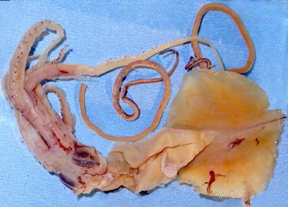
Introduction
Nesis (1977) described M. psychrophila from Antarctic waters on the basis of four specimens, the largest of which was a nearly mature male of 143 mm ML. Critical details of the photophore pattern remain unknown.
Diagnosis
A mastigoteuthid of the M. agassizii group ...
- without large, lateral pegs on outer rings of club suckers.
- with large tragus and distinct antitragus on funnel locking-apparatus.
Characteristics
- Tentacles
- Club suckers small (diameter ca. 0.15 mm) (Nesis, 1977).
- Club suckers circular without large inward directed knobs; outer rings with 12-15 short, blunt-rounded knobs around entire perimeter (Nesis, 1977).
- Funnel locking apparatus
- Funnel locking-apparatus with well developed tragus and distinct antitragus (Nesis, 1977).
- Photophores
- Mantle, head, arms and fins (dorsal and ventral surfaces) with small integumental photophores (Nesis, 1977).
- Closely spaced photophores in the transverse surface on arms IV suggest that more than two series exist.


Figure. Club suckers of M. psychrophila. Left - Oral view of one sucker, holotype. Drawing from Nesis (1977). Right - Oral view of a portion of the club, holotype. Note the interspersed small suckers which may regeneration stages of previously lost suckers. Photograph by M. Vecchione.
Scanning electron micrographs of the suckers can be found here.

Figure. Frontal view of the funnel locking-apparatus of M. psychrophila, holotype. Drawing from Nesis (1977).


Figure. Funnel/mantle locking-apparatus of M. psychrophila. Left - Frontal view of funnel component, NMNH 817317, methylene blue stain. Left-center - Funnel component cut along midline and turned back to show longitudinal sections of the lock, NMNH 817317, methylene blue stain. Note the strongly undercut antitragus. Right-center - Frontal view of the mantle component, NMNH 817317. Photographs by R. Young. Right - Frontal view of the funnel component, holotype. Photograph by M. Vecchione.


Figure. The specimens examined had lost most of the integument but a few photophores remained. Left - Ventral view of the anterior tip of the head and base of arms IV. Square shows a patch of remaining photophores that is enlarged at center. Note also the closely spaced patch of photophores on the left arm IV at the top of the photograph. Center - Enlargement of integumental photophores. Photographs by R. Young. Right - Aboral view of arm IV photophores showing closely spaced photophores, holotype. Photograph by M. Vecchione.
Comments
More details of the description can be found here.
M. psychrophila is very similar to other species in the M. agassizii species-group. Its most distinctive features are the shape of the funnel locking-apparatus and the apparent lack of tentacular suckers with pronounced lateral pegs on the outer sucker rings. Unfortunately the pattern of photophores on arms IV is unknown other than that it appears to differ from most members of the group in having more than two series of photophores.
Distribution
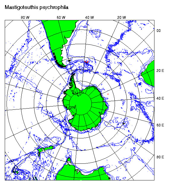
Type locality: 59°26'S, 158°36'E, Antarctic waters south of eastern Australia in an open midwater trawl that fished mostly at 500 m depth at night. Nesis (1977) also reports a record in the same region at 55°S and in South Atlantic Antarctic waters at 57°S, 26°W. He states that they were taken from water of Antarctic and Subantarctic structure. The NMNH 817317 squid came from 58°S, 77°W just into the Pacific Antarctic waters. The NMNH 884836 squid came from 62°50'S and 114°30'W. The distribution appears to be circumantarctic occurring both north and south of the Antarctic Convergence (Nesis, 1977). Additional records are found at the British Antarctic Survey .
References
Nesis, K.N. 1977. Mastigoteuthis psychrophila sp. n. (Cephalopoda, Mastigoteuthidae) from the Southern Ocean. Zoologichesky Zhurnal, 65(6):835-842.
Title Illustrations
| Scientific Name | Mastigoteuthis psychrophila |
|---|---|
| Location | 59?26'S, 158?36'E |
| Specimen Condition | Dead Specimen |
| Sex | Male |
| View | Ventral |
| Size | 84 mm ML |
| Type | Holotype |
| Copyright | © |
About This Page
Richard E. Young
Dept of Oceanography
University of Hawaii
Honolulu, Hawaii 96822
USA
National Marine Fisheries Service
Systematics Laboratory
National Museum of Natural History
Washington, D. C. 20560
USA
Page copyright © 2004 Richard E. Young and
- First online 05 March 2004
Citing this page:
Young, Richard E. and Vecchione, Michael. 2004. Mastigoteuthis psychrophila . Version 05 March 2004 (under construction). http://tolweb.org/Mastigoteuthis_psychrophila/19522/2004.03.05 in The Tree of Life Web Project, http://tolweb.org/

