


Megalocranchia
Richard E. Young and Katharina M. Mangold (1922-2003)

Megalocranchia contains four recognized species. Voss, et al. (1992), however, suggest that six species exist.
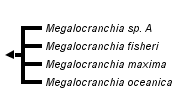


This tree diagram shows the relationships between several groups of organisms.
The root of the current tree connects the organisms featured in this tree to their containing group and the rest of the Tree of Life. The basal branching point in the tree represents the ancestor of the other groups in the tree. This ancestor diversified over time into several descendent subgroups, which are represented as internal nodes and terminal taxa to the right.

You can click on the root to travel down the Tree of Life all the way to the root of all Life, and you can click on the names of descendent subgroups to travel up the Tree of Life all the way to individual species.
For more information on ToL tree formatting, please see Interpreting the Tree or Classification. To learn more about phylogenetic trees, please visit our Phylogenetic Biology pages.
close boxIntroduction
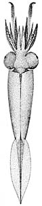
Some species of Megalocranchia are very large and can reach 1800 mm ML, (Tsuchiya and Okutani, 1993). The vertical distribution is best known in M. fisheri. The paralarval stage, which reaches 40-50 mm ML, is spent in near-surface waters. Larger individuals occupy mesopelagic depths during the day and migrate into near-surface waters at night (Young, 1978).

Figure. Side view of earyl juvenile M. fisheri. Photograph by R. Young.
Brief diagnosis:
A taoniin ...
- with photophores on the digestive gland.
Characteristics
- Tentacles
- Tentacular clubs with suckers only.
- Tentacular stalk with two series of suckers and pads in mid-third, then four series to carpal group.
- Funnel
- Funnel valve present.
- Funnel organ: Dorsal pad with two triangular flaps, no papillae.
- Mantle
- Tubercules absent at funnel-mantle fusion.
- Paralarvae (to ca. 50 mm ML) with thick gelatinous dermis on mantle.*
- Fin
- Anterior 10-15% of fin inserts on mantle (in subadults).**
- Anterior 10-15% of fin inserts on mantle (in subadults).**
- Photophores
- Photophores present on digestive gland.*
- Photophores present on tips of arms I-III or only II or only III of adult females.Click on an image to view larger version & data in a new window

Figure. Visceral photophores from a subadult, M. fisheri, off Hawaii. The visceral photophores contain four reflecting cups (green). The intestine, partially covered with red pigment, is seen here passing between photophores of either side. The barely-discernable digestive gland is the fuzzy central dark region. Photograph by R. E. Young.
*Within family, unique to this genus.
**Within family, unique degree of mantle attachment. Most genera lack a mantle attachment but two have more than 30% of fin attached to the mantle(Egea, Teuthowenia) rather than to the gladius.
Comments
Characteristics are from Voss (1980).
Life History
Paralarvae of M. fisheri from Hawaiian waters have been identified. The large posterodorsal mantle chromatophore seen in both stages is distinctive among cranchiid paralarvae from these waters. What appears to be an unusually long club may actually be a combination of the club with a large number of carpal suckers on the distal stalk.
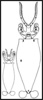
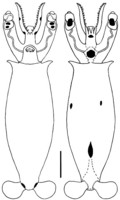

Figure. Paralarvae of M. fisheri, Hawaiian waters. Thumbnail (far left) - Illustration shows relative sizes of the two paralarvae. Left - Ventral and dorsal views of a 4.9 mm ML paralarva. Right - Ventral and dorsal views of a 14.5 mm ML paralarva. The scale bars are 1 mm. Drawings by R. Young.
References
Tsuchiya, K. and T. Okutani. 1993. Rare and interesting squids in Japan -X. Recent occurrences of big squids from Okinawa. Venus 52: 299-311.
Voss, N. A. (1980). A generic revision of the Cranchiidae (Cephalopoda; Oegopsida). Bull. Mar. Sci., 30: 365-412.
Voss N. A., S. J. Stephen and Zh. Dong 1992. Family Cranchiidae Prosch, 1849. Smithson. Contr. Zool., 513: 187-210.
Young, R. E. 1978. Vertical distribution and photosensitive vesicles of pelagic cephalopods from Hawaiian waters. Fish. Bull. 76: 583-615.
Title Illustrations

| Scientific Name | Megalocranchia fisheri |
|---|---|
| Location | off Hawaii |
| Specimen Condition | Dead Specimen |
| Sex | Female |
| Size | large, 2.7 m total length |
| ToL Image Use |
 This media file is licensed under the Creative Commons Attribution-NonCommercial License - Version 3.0. This media file is licensed under the Creative Commons Attribution-NonCommercial License - Version 3.0.
|
| Copyright |
© Richard E. Young

|

| Scientific Name | Megalocranchia |
|---|---|
| Reference | Voss, N. A. (1980). A generic revision of the Cranchiidae (Cephalopoda; Oegopsida). Bull. Mar. Sci., 30: 365-412. p. 399, Fig. 12a |
| Copyright | © 1980 Bulletin of Marine Science |
About This Page
Drawings from Voss (1980) printed with the Permission of the Bulletin of Marine Science.
Richard E. Young
University of Hawaii, Honolulu, HI, USA
Katharina M. Mangold (1922-2003)
Laboratoire Arago, Banyuls-Sur-Mer, France
Page copyright © 1996 Richard E. Young and Katharina M. Mangold (1922-2003)
 Page: Tree of Life
Megalocranchia .
Authored by
Richard E. Young and Katharina M. Mangold (1922-2003).
The TEXT of this page is licensed under the
Creative Commons Attribution-NonCommercial License - Version 3.0. Note that images and other media
featured on this page are each governed by their own license, and they may or may not be available
for reuse. Click on an image or a media link to access the media data window, which provides the
relevant licensing information. For the general terms and conditions of ToL material reuse and
redistribution, please see the Tree of Life Copyright
Policies.
Page: Tree of Life
Megalocranchia .
Authored by
Richard E. Young and Katharina M. Mangold (1922-2003).
The TEXT of this page is licensed under the
Creative Commons Attribution-NonCommercial License - Version 3.0. Note that images and other media
featured on this page are each governed by their own license, and they may or may not be available
for reuse. Click on an image or a media link to access the media data window, which provides the
relevant licensing information. For the general terms and conditions of ToL material reuse and
redistribution, please see the Tree of Life Copyright
Policies.
- Content changed 15 July 2008
Citing this page:
Young, Richard E. and Mangold (1922-2003), Katharina M. 2008. Megalocranchia . Version 15 July 2008 (under construction). http://tolweb.org/Megalocranchia/19562/2008.07.15 in The Tree of Life Web Project, http://tolweb.org/

