- Photophores (head)
- Compound photophores (Type 1b):
- Basal Arm IV Row with 3 photophores. Medial photophore separated by 1 photophore from anterior photophore of Midline Series
- Midline Series with 3 photophores.
- Right Eyelid Series with 17 photophores.
- Left Eyelid Series with 6 photophores.
- 2° Right Eyelid Series with 8 photophores.
- 2° Left Eyelid Series with 7 photophores.
- Ventral Matrix with 36 photophores including an asymmetrically placed dangling, rogue photophore (colored red in illustration), and 9 longitudinal (blue) series. Attachment to 2° Eyelid Series at 2° eyelid photophores no. 2 either side.
- Ventral Matrix photophores do not align in transverse rows.
- Right basal series absent.
- Left basal series absent.
- Basal row with 9 photophores and one sawtooth (note that two of the sawtooth photophores align with the rogue (red) photophore..
- Right Accessory Series with 2 photophores (see below).
- Left Accessory Series with 3 photophores (see below).
 Click on an image to view larger version & data in a new window
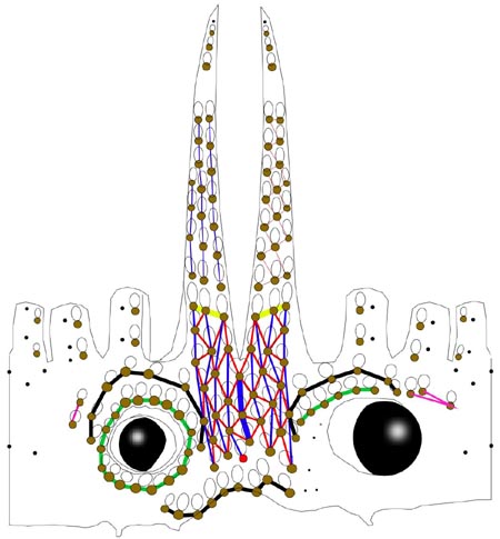
Figure. 360° view of arrangement of photophores on the head and arm bases of Histioteuthis sp. A. Dorsal, lateral and ventral views combined in the illustration; the left and right edges of the illustration are the mid-dorsal line of the head. Drawing by R. Young.
- Right Basal Series absent.
- Left Basal Series apparently absent.
- Basal Row with 9 photophores and one sawtooth (note that two of the sawtooth photophores align with the rogue (red) photophore.
- Right Accessory Series with 2 photophores (see below).
- Left Accessory Series with 3 photophores (see below).
- Simple photophores (round, black dots):
- Arrangement as illustrated above.
- Large, simple photophores on dorsal surface of head absent.
- Small, simple photophores not easy to recognize and may be variable.
- Compound photophores (Type 1b):
- Compound photophores (arms)
- Arms IV with 3 longitudinal series of photophores on arm base (photograph below, drawing above).
- Single series of compound photophores on ends of arms IV (at least) in immatures, distinctly separated from regular arm series (photograph below).
- Arms III with one primary longitudinal series and without secondary series at bases.
- Arms II with one primary longitudinal series and without secondary series at bases.
- Arms I with one primary longitudinal series and without secondary series at bases.
Click on an image to view larger version & data in a new window
Figure. Ventral view of arm IV of Histioteuthis sp. A, 25 mm ML, Hawaii. Photograph by R. Young.
- Compound photophores (mantle)
- Photophores on anterior half of ventral mantle of uniform size.
- Photophores align in transverse rows.
- Photophores from the anterior region of the ventral mantle measure 0.95-1.1 mm across the pigmented cup in a 40 mm ML squid (i.e., 2.4-2.8 % of ML).
Click on an image to view larger version & data in a new window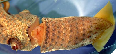
Figure. Ventral view of the mantle of Histioteuthis sp. A, 25 mm ML, Hawaii. Photograph by R. Young.
- Arms
- Arm suckers with smooth inner rings. Click on an image to view larger version & data in a new window
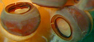
Figure. Oblique oral view of suckers of arm II of Histioteuthis sp. A, 41 mm ML, Hawaii. Photograph by R. Young.
- Tentacles
- Club with about 7 irregular longitudinal sucker series.
- Suckers across manus of nearly equal size; medial suckers only slightly enlarged.
- Suckers with long, ~15-20 slender teeth on distal half of inner ring.
- Suckers with broad outer rings.
- Suckers of two ventral-marginal series without noticeably asymmetrical outer rings (photographs below).
- Web and buccal crown
- Inner web virtually absent; outer web low.
- Buccal membrane with XXXX
- Head
- Two well-developed occipital folds on either side of head.
- Funnel organ
- Inverted V-shaped dorsal organ without noticeable sculpture.
- Fins
- Fin length nearly half the mantle length.
- Gladius
- Not examined
- Spermatophore
- Unknown
- Hectocotylus
- Unknown
Photographs showing photophore patterns can be seen here.
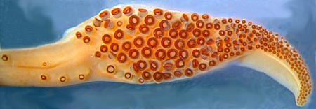
Figure. Oral view of club of Histioteuthis sp. A, 41 mm ML, Hawaii. Photograph by R. Young.
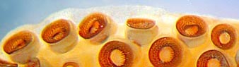
Figure. Oral view of two ventral-marginal sucker series on the manus of the club of Histioteuthis sp. A, 41 mm ML, enlargement from club illustrated above. Photograph by R. Young.
Compare with scanning electron micrographs of the suckers.
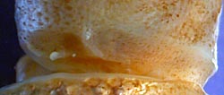
Figure. Lateral view of occipital folds and olfactory organ of Histioteuthis sp. A, 25 mm ML, Hawaii. Photograph by R. Young.
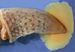
Figure. Ventral view of mantle and fins of Histioteuthis sp. A, 41 mm ML, Hawaii. Photograph by R. Young.
Comments
This species is very similar to the Atlantic Ocean Histioteuthis celetaria. Only two feature clearly separate them at present: the absence of asymmetrical outer rings in the ventral marginal suckers of the club in Histioteuthis sp. A and it's North Pacific habitat.


