Note

Histioteuthis sp. A: Sucker structure

Richard E. Young and Michael Vecchione

Suckers of two of  paratypes were examined with the scanning electron microscope (SEM). Suckers were prepared by dehydration in absolute ethanol then air drying.
  1. Arm suckers
  2. Click on an image to view larger version & data in a new window
    Click on an image to view larger version & data in a new window

    Figure. Oral view of arm suckers of Histioteuthis sp. A, 40 mm ML, Hawaiian waters (21°20'N, 158°20'W). Left - Arm II, sucker V8 (sucker 8 of ventral series). Right - Arm III, sucker V4. Photographs by R. Young.

    Click on an image to view larger version & data in a new window
    Click on an image to view larger version & data in a new window

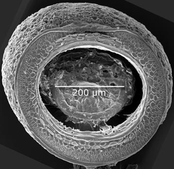
    Figure. Oral view of arm IV sucker V4 of Histioteuthis sp. A, same specimen as above. Photograph by R. Young.

  3. Tentacular club suckers
  4. Click on an image to view larger version & data in a new window
    Click on an image to view larger version & data in a new window

    Figure. Oral views of club suckers of Histioteuthis sp. A, same specimen as above, from the two ventral-most series on the club manus. Their positions on the club are indicated on the photograph below. The smaller sucker to the left is from the submarginal series and the sucker to the right is from the marginal series. Photographs by R. Young.

    Click on an image to view larger version & data in a new window
    Click on an image to view larger version & data in a new window

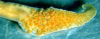
    Figure. Oral view of the club of Histioteuthis sp. A from which the suckers for the adjacent SEM photographs were taken. The bottom circle indicates the location of the two suckers viewed above. The two upper circles indicate the location of the suckers viewed below. Suckers from the two marginal series had been removed prior to taking the photograph. Photograph by R. Young.

    Click on an image to view larger version & data in a new window
    Click on an image to view larger version & data in a new window

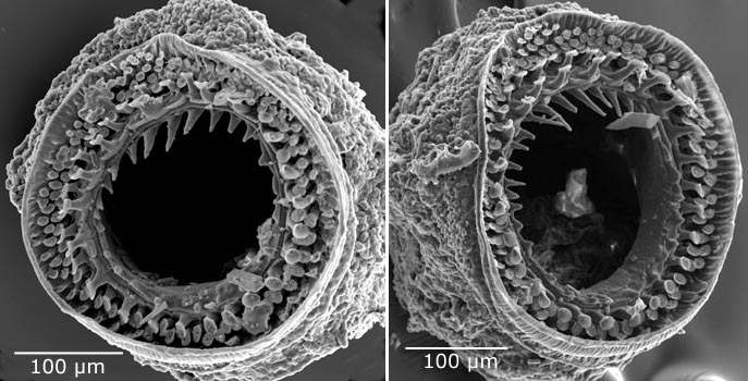
    Figure. Oral views of club suckers from the central region of the manus of Histioteuthis sp. A, same specimen as previous photographs. Their positions on the club are indicated on the photograph above.. Photographs by R. Young.

    Click on an image to view larger version & data in a new window
    Click on an image to view larger version & data in a new window

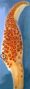
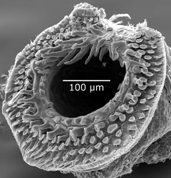
    Figure. Oral view of sucker from ventral-marginal series of the club of Histioteuthis sp. A, 41 mm ML, same capture locality as the previous specimen. Left - oral view of the club with the sucker encircled that was examined with SEM. Right - SEM photograph of the encircled sucker. Sucker was damaged distally during preparation. Photographs by R. Young.


About This Page


University of Hawaii, Honolulu, HI, USA


National Museum of Natural History, Washington, D. C. , USA

Page: Tree of Life Histioteuthis sp. A: Sucker structure Authored by Richard E. Young and Michael Vecchione. The TEXT of this page is licensed under the Creative Commons Attribution-NonCommercial License - Version 3.0. Note that images and other media featured on this page are each governed by their own license, and they may or may not be available for reuse. Click on an image or a media link to access the media data window, which provides the relevant licensing information. For the general terms and conditions of ToL material reuse and redistribution, please see the Tree of Life Copyright Policies.

close box

This page is a note that is attached to a leaf of the Tree of Life.

ToL notes provide brief accounts of characteristics, short summaries, commentaries, media files, taxonomic information, or identification tools for a given group of organisms.

For a more detailed explanation of the different ToL page types, have a look at the Structure of the Tree of Life page.

close box

Histioteuthis sp. A

Page Content

articles & notes

collections

people

Explore Other Groups

random page

  go to the Tree of Life home page
top