I. External structure

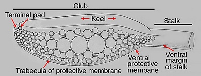
Figure. Tentacular club of Sepia Officinalis, Oral view. Drawing from Naef 1921-23.
Features:
- Carpus: Absent.
- Club divisions: Primary club (not divided into manus and dactylus), terminal pad.
- Club shape: Expanded, often short; may or may not extend distally with gradually narrowing region; generally with distal dorsal curvature.
- Sucker series: 4-8 very irregular longitudinal series; with mostly 4-8 suckers in irregular transverse series (Naef, 1921-23).
- Trabeculate, protective membranes: Trabeculae of dorsal membrane cannot be detected (see section III below); trabeculae of ventral membrane difficult to detect; dorsal and ventral membranes well developed over full length of club; both membranes join or nearly join at proximal end of club; dorsal protective membrane larger than ventral membrane.
- Oral margins of stalk: Present, but very reduced and without trabeculae (Naef, 1921-23). (1) When protective membranes of club do not join proximally, oral margins of stalk abut/join protective membranes of club (see drawing of S. officinalis above). (2) When protective membranes joined proximally, oral margins of stalk may fuse with one another prior to abutting/joining protective membranes of club (see S. latimanus below).
- Aboral margin of stalk: Present but low and rounded (Naef, 1921-23); not apparent in our specimen.
- Keel: Extends full length of club, or further. Keel positioned dorsally throughout its length and adjacent to the dorsal protective membrane.
- Terminal pad: Present but with few suckers.
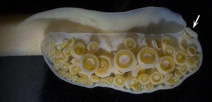
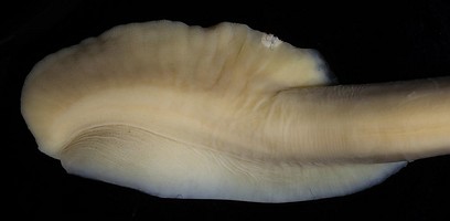
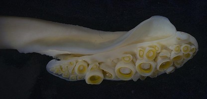
Figure. Sepia latimanus, male, 145 mm ML. Top - Oral view of club. Arrow points to the terminal pad with its single sucker. Middle - Aboral view. Note that the distal dorsal curvature of the club is apparent only in the aboral view. Bottom - Dorsal view.
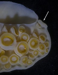
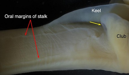
Figure. Sepia Latimanus, 145 mm ML. Left - Enlarged oral view of the club tip showing the Terminal pad (arrow). Right - Oral view of the club stalk and its junction with the club. The proximal end of the club has been folded forward to show the single membrane (arrow) extending from the club protective membrane onto the oral face of the stalk where the membrane divides to form the two, small, oral margins of the stalk.
II. Internal structure: Skin, suckers removed
- Dorsal view: The large central suckers (black arrows, below), and the smaller more marginal sucker stalks, are bathed along their dorsal and lateral margins in sea water as they emerge from the club core; a very peculiar arrangement. The dorsal edges of the central sucker-stalks, at their origin, are positioned further dorsally than those of the more dorsal suckers.
- Aboral view: Ventromarginal and some near ventromarginal sucker-stalks are very long; they appear in the photograph below to originate from near the midline of the club core and to, virtually, extend to the ventral margin of club leaving room for only a very short, free, protective membrane. These stalks seem to be of slightly varying widths, a result of the slightly varying positions of the ventral-marginal suckers.
- Oral view: Stalks of large central sucker, and more ventral suckers, strongly tilted relative to those of the more dorsal suckers indicating the peculiar arrangement in sucker attachment. Musculature between suckers, suckers and trabeculae, and trabeculae complex and poorly understood.

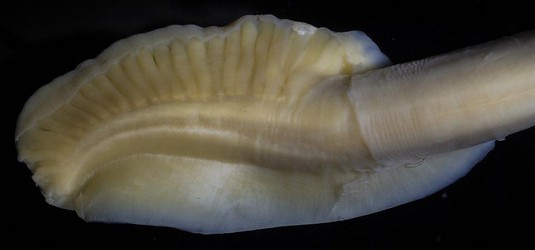
Figure. S. latimanus, 145 mm ML. Top - Dorsal view, with protective membrane folded orally, that reveals, in the large space open to sea water, the sucker stalks of the large central suckers (black arrows), along with those of smaller suckers, arising directly from the club core. This is an apparently unique method of bathing the sucker stalks in sea water. Bottom - Aboral view with skin removed showing the long stalks of lateral and near-lateral suckers.
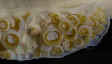
Figure. Sepia Latimanus, 145 mm ML. Oral view of posterior region of club with some suckers removed showing the club surface beneath the suckers. Note that trabeculae are visible in the ventral membrane at the right of the image. Red dots - Indicate stalks of removed suckers.
- Organization: Strong asymmetry with very long stalks for ventral suckers grading to small stalks for dorsal suckers.
- Dorsal trabeculate protective membrane: Forms broad, solid, thick, muscular layer (presumably through fusion of trabeculae); Dorsomarginal sucker-stalks fused along one side with this muscular membrane.
- Ventral trabeculate protective membrane: Narrow; membrane and trabeculae attach primarily to distal parts of ventromarginal sucker-stalks.
- Core shape: Circular with slightly flattened "top"; appears tilted toward ventral margin compared to that of tentacular stalk.
- Keel: Attached dorsally throughout its length and adjacent to dorsal protective membrane
- Ventromarginal and ventral submarginal sucker-stalks fused along their adjacent margins.
- Water canals. Present but unique. Between keel and dorsal protective membrane sucker stalks exposed to sea water via canals (i.e., spaces) extending well past exposed sucker-stalks to dorsal surfaces of ventral submarginal sucker-stalks. However, the separate incursion of sea-water spaces on sides of exposed sucker stalks do not appear to connect with one another or with the oral surface of the club.


Figure. Sepia sp. Left - Cross-section through the middle of the club showing the basic structure of the club core, and the attachment of the keel, sucker stalks and trabeculae of the ventral protective membrane. Most of the core outside the axial nerve cord is composed of muscles. Dots on drawing areas indicate primarily longitudinal muscles. Other colors and fill-lines indicate more complex musculature. The red dot, on the aboral side of the nerve cord, is the major artery of the club; the blue dot, aboral to the nerve cord, is the major vein of the club. The drawing by R. Young is a compilation taken from histological cross-sections. Right - Cross-sectional cut with a scapel of the tentacular stalk just proximal to the club, for comparison with the club. Note the major vein that drains the club is located in the oral-ventral corner of the stalk. .


