
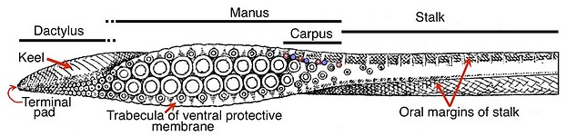
Figure. Tentacular club of Ommastrephes bartramii, Oral view. Drawings from Naef 1921-23. Not all basic clubs, however, are divided into dactylus, manus and carpus.
The basic type of tentacular club for the Decapodiformes is made up SOME or ALL of the following parts:
- Terminal pad - Lies at anterior end of club (i.e., distal to the dactylus (when present)); consists of 1-20 or so, smooth ringed suckers. All suckers are often tightly packed together in an oval or slightly elongate shape and have short, approximately equal-sized stalks. A flap of skin curves over the distal edges of the pad. The aboral side of the pad contains what appears to be a thick dermis of dense connective tissue not found elsewhere on the club.
The function of the terminal pad as a distal locking-mechanism is known, from experimental evidence, only in loliginids (loliginids lack a carpus): "As the tentacles elongate, they twist so that the suckers face outward and the tips of the clubs remain attached" (Keir and van Leeuwen, 1997, an experimental analysis of Loligo pealei attacking a tethered shrimp). In oegopsids the terminal pad seems to act as a distal locking-mechanism to help hold the clubs together, at least, when not in use (see photographs below). The locking mechanism differs from that of the carpal locking mechanism as the pad has no knobs for the suckers to lock onto. The mechanism seems to consist of the suckers of one pad locking onto the aboral surface of the pad of the opposite club (see Figure below). Evidence for the use of the terminal pad as a locking mechanism during an attack on prey is suggestive in oegopsids (see Dosidicus gigas, Behavior).
Click on an image to view larger version & data in a new window
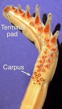
Figure. Left - Two photos of Onychoteuthis cf. equimanus, taken in situ in the open ocean, show the clubs in two different states. The squid at the left is in a dorsal arm-curl posture with its tentacles contracted and its cubs tightly locked together. The enlargement of the tip of the clasped tentacles shows the terminal pad of one (yellow arrow) seemingly attached to the aboral surface of the other. The squid at the right has started to extend its tentacles. The enlargement of the tentacles shows that the clubs are separated except at the carpus (upper yellow arrow) and the terminal pad (lower arrow). Please click on the photograph to view the larger image. Photographs by Jeffrey Milisen. Right - Onychoteuthis compacta, oral view of a preserved club showing structure. Most onychoteuthids have no dactylus.
- Dactylus - Narrow region that grades proximately into the manus and ends distally at the terminal pad. This region is narrower than the manus and strongly asymmetrical between dorsal to ventral sides with differences in the lengths of the sucker stalks and widths of the trabeculate protective membranes.
- Manus - The longer and, generally, wider portion of the club. Width usually expanded, relative to that of the tentacular stalk, by four series of suckers with the lateral suckers set on longer stalks and by wide trabeculate protective membranes. The manus, typically appears somewhat bilaterally symmetrical with (1) suckers of the dorsal-medial series of equal size as their partners of the ventral-medial series, and the same for suckers of the lateral two series and (2) similarly sized trabeculate protective membranes on either side of the manus. The four suckers series, however, are not aligned at right angles to the axis of the club, as would be required for bilateral symmetry but are aligned at both oblique and acute angles. Bilateral symmetry is also often defeated by small differences in trabeculae of the dorsal and ventral protective membranes, and by the positions of the keel and carpal suckers and knobs.
- Carpus - Structure consisting of suckers and knobs that lies at the proximal end of club and, often, overlaps the manus along its dorsal border, and may extend well down the oral surface of the tentacular stalk. This is the proximal locking-mechanism of the club where the muscular, carpal knobs, of one tentacle lock onto the horny, smooth-ringed, carpal suckers of the opposite tentacle.
- Trabeculate protective membranes - Membranes along the dorsal and ventral margins of the club that have muscular supporting pillars, the trabeculae. The membranes may extend the length of the club or be greatly restricted or elaborated. The proximal ends of the membranes may be continuous with the oral margins of the stalk.
- Keel - Muscular vane that may extend the length of the club or be restricted to the distal region or absent. The proximal end of the keel may be continuous with the aboral margin of the stalk.
- Water canals - Spaces within the club that, in life, are open to sea water and that pass beneath much of the integument and surface musculature of the oral face of the club. Water spaces commonly surround the sucker stalks on some clubs but they do not form canals. Presumably canals increase flexibility in altering the shape of the club.
What are the basal taxa?
Within the Decapodiformes there are 7 major monophyletic groups: Sepiidae, Idiosepiidae, Sepiolida, Spirulidae, Myopsida, Bathyteuthoidea, Oegopsida. The relationships between these taxa has not been clearly resolved (see Lindgren, 2010, Lindgren, et al., 2012). Of these taxa three are families, three are composed of two families and one, Oegopsida of 24 families. We have picked one or two representative from each of 6 of the major taxa based on assumed basal position within their group or availability. For the Oegopsida we have picked representatives from two families, Thysanoteuthidae and Ommastrephidae, based on the suspected early derivation, as sister families, within the Order (Lindgren, et al., 2012). Our basal clubs are from:
- Myopsida - Loligo forbesi (information from Naef, 1921-23) and Sepioteuthis sp.
- Oegopsida - Thysanoteuthis rhombus (Thysanoteuthidae), and Ommastrephes bartramii and Sthenoteuthis oualaniensis (Ommastrephidae).
- Sepiidae - Sepia officinalis and Sepia latimanus.
- Spirulidae - Spirula spirula.
- Sepiolida - Rossia pacifica (Sepiolidae).
- Bathyteuthoidea - Chtenopteryx sp.
- Idiosepiidae - Idiosepius sp.
Origin of the tentacular clubs
The tentacles are considered to be modifications of the 4th pair of arms of an ancestor with 10 arms (Naef, 1921-23). Therefore the structure of the arm should provide information on the original structure of the tentacular club. The ancestral decapods are thought to have arms with a single series of suckers from which were derived, in succession, arms bearing two series, then four series then more than four series (Naef, 1921-23). We therefore take as a typical example an arm that of Ommastrephes bartramii, a member of one of our basal decapodiform taxa which bears arm suckers in two series (no decapodiform has a single series) and a species that we have histological sections from.
- Comparison of the arm and club of O. bartramii.
- Suckers of arm more widely separated from adjacent suckers than those of club.
- Ventral and dorsal arm-sucker series are offset making the "transverse" sucker rows, consisting of two suckers, oblique to the arm axis; the "transverse" suckers rows of the club, consisting of four suckers, show the same oblique orientation.
- Arm trabeculae attach to sucker stalks; club trabeculae attach partly to lateral sucker stalks and partly to club core
- Arm with one trabecula per sucker; club with two per lateral sucker on the manus but one per lateral sucker on the dactylus.
Click on an image to view larger version & data in a new window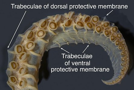
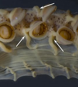
Figure. Ommastrephes bartramii, immature female, 155 mm ML. Left - Oral view of arm III; note the very long but highly folded ventral trabeculae compared to the short dorsal trabeculae (arrows). Right - Magnification of the mid-portion of the adjacent photograph showing attachment of trabeculae to the sucker stalks (arrows).
Click on an image to view larger version & data in a new window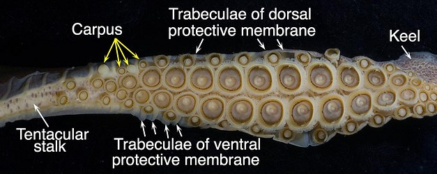
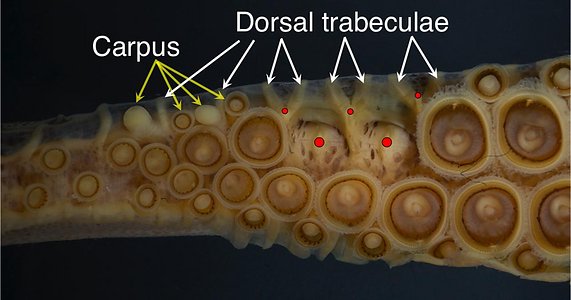
Figure. Ommastrephes bartramii, 155 mm ML. Top - Oral view of the tentacular club. Most of the dactylus is not shown in the image as it was damaged and in the process of regeneration. Bottom - Oral view of the proximal region of the same club with some suckers removed. Red dots - Indicate where the necks of the sucker stalks were cut to remove the suckers.
- Suckers of arm more widely separated from adjacent suckers than those of club.
- Comparison of cross-sections from the arm and club manus of O. bartramii.
- Arm suckers at least as close to the midline of the arm as the medial club suckers are to the midline of the manus; however, within each sucker series (i.e., arm suckers vs medial club suckers) the arm suckers are much further apart.
- Arm suckers with stalks intermediate in length between those of the lateral and medial suckers of the club manus.
- Structure of arm core arm very similar to that of manus.
- Trabeculae of arms more fully attached to sucker bases than those of club manus.
- Attachment of suckers to core similar in arms and manus.
Click on an image to view larger version & data in a new window
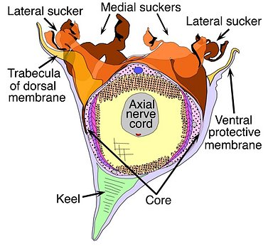
Figure. Ommastrephes bartramii. Left - Arm III cross-sections (proximal view from compilation of sections); 113 mm ML. . Right - Club cross-sections (xxx view from compilation of sections); 120 mm ML.
- Arm suckers at least as close to the midline of the arm as the medial club suckers are to the midline of the manus; however, within each sucker series (i.e., arm suckers vs medial club suckers) the arm suckers are much further apart.


