I. External structure

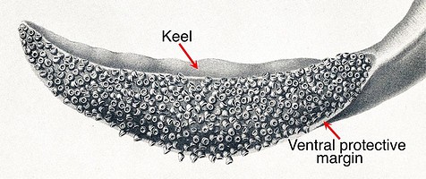
Figure. Tentacular club of Spirula spirula, Oral view. Drawing from Chun, 1910.
Features:
- Carpus: Absent.
- Club divisions: None (not divided into manus and dactylus, presence of terminal pad uncertain).
- Club shape: Expanded slightly along ventral side; tapers to a rounded distal end; exhibits a general dorsal curve which is accentuated near tip.
- Sucker series: Numerous, irregular longitudinal series (approximately 8-16 series -Naef, 1921-23).
- Trabeculate, protective membranes: Absent; ventral margin marked by rounded edge; dorsal margin not found in our specimens. Position of edges at proximal end of club uncertain.
- Oral margins of stalk: Not found in specimens examined.
- Aboral margin of stalk: Not found in specimens examined.
- Keel: Present on distal two thirds of club; positioned dorsally throughout its length.
- Terminal pad: Uncertain. The tip of the club makes a tight turn dorsally and carries distal flap, both common in terminal pads but more clubs need to be examined.
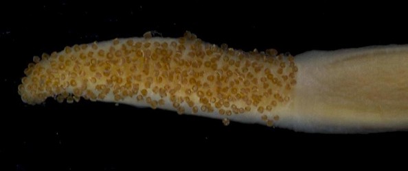
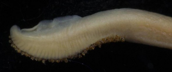
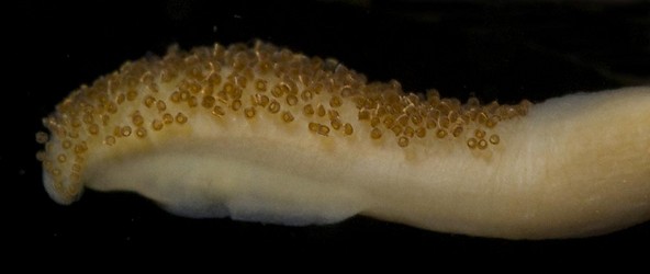
Figure. Spirula spirula, Female, 32 mm ML, tentacular club. Top - Oral view. Middle - Aboral view. Note the dorsal curvature of the club core. Bottom - Dorsal view showing more clearly where the keel attaches.

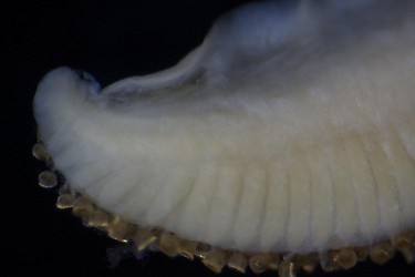
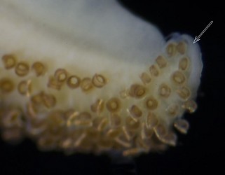
Figure. Spirula spirula , same tentacular club. Top - Ventral view, with light methylene blue stain showing the ventral margin (arrow); no dorsal margin was detected in the staining. Bottom left - Aboral view of distal half of club showing the long stalks of the ventral marginal suckers in more detail. Photomicrograph. Bottom right - Oral-distal view of the possible terminal pad. Arrow points to what could be a distal flap of a terminal pad. Photomicrograph.
II. Internal structure: Cross-section at mid-club
- Suckers have very long necks extending from their stalks.
- Each sucker stalk basically has an inner, ovoid region covered by a coat of intercrossing fibers that mostly pass through a thick outer layer of the core (pink in drawing) to attach to the underlying layer of the core.
- The mid-club has a ventral bias with the stalks becoming progressively longer ventrally.
- No trabeculate membranes are present although slight margins exist on either side of the club (Naef, 1921-23).
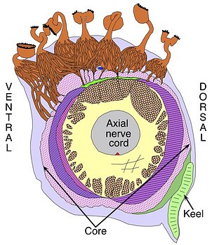
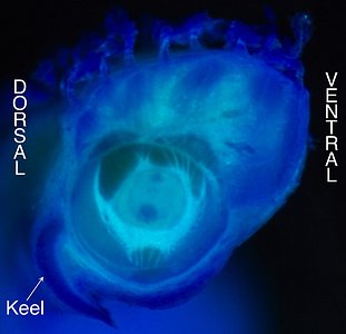
Figure. Spirula spirula, Left - Female, 37 mm ML. Cross-section through mid-club showing the basic structure of the club core, and the attachment of the keel and sucker stalks. Most of the core outside the axial nerve cord is composed of muscles. Dots on drawing areas indicate primarily longitudinal muscles. Other colors and fill-lines indicate more complex musculature. The red dot, on the aboral side of the nerve cord, is the major artery of the club; the blue dot, oral to the nerve cord, is the major vein of the club. More of the outer layer of the intercrossing fibers of the stalk reach through the pink layer than shown (this was done prevent obscuring the large pink layer). The drawing by R. Young is a compilation taken from histological cross-sections. Right - Mature male, 35 mm ML. Single cut, using scalpel, through middle of club and lightly stained with methylene blue to show attachment of keel. Also a hint of the outer and inner portions of the sucker stalks are apparent.


