I. External structure

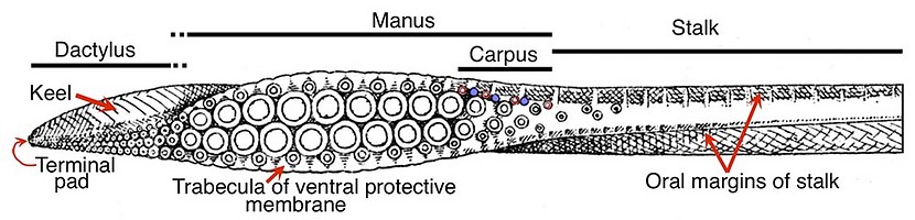
Figure. Tentacular club of Ommastrephes bartramii, Oral view. Drawings from Naef 1921-23.
Features:
- Carpus: Present, but only in Ommastrephinae within Ommastrephidae. Carpus seen above consists of three pads (blue) and four suckers (red) along the dorsal-proximal margin of the manus. On the opposite club there would be four pads and three suckers.
- Club divisions: Carpus, manus, dactylus and terminal pad.
- Club shape: Expanded manus with narrow dactylus generally having a dorsal curvature (latter not seen in drawing above).
- Sucker series: Four, regular, longitudinal series except for few suckers at at proximal end of club. Four suckers comprising a transverse row are arranged at oblique and acute angles to the axis of the club rather than at a right angle.
- Trabeculate, protective membranes: Two trabeculae per lateral sucker on manus, one per lateral sucker elsewhere; dorsal membrane low to virtually absent on distal half of dactylus; dorsal and ventral membranes broadly separated at proximal end of club.
- Oral margins of stalk: Present; prominent; with distinct trabeculae; located at lateral sides of oral surface of stalk; continuous with trabeculate protective membranes of club; extend full length of tentacular stalk.
- Aboral margin of stalk: Present; prominent, much larger than oral margins; positioned on aboral-dorsal corner of stalk; extends distally to level of mid-manus; continuous in position and structure (i.e., muscular) with keel; extends proximally full length of tentaclular stalk.
- Keel: Extends from dorsal position on dactylus to middle of manus where it becomes low and aboral in position; then continues proximally on stlak as a tall aboral margin.
- Terminal pad present; usually distinct.
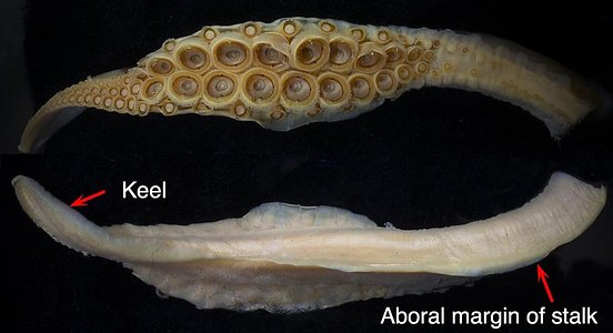
Figure. Sthenoteuthis oualaniensis, immature female, 142 mm ML. Oral (top) and Aboral (bottom) views of the club. Note that the keel is continuous with the aboral margin of the stalk (see II and III below also) and that the trabeculate membranes are present on the oral margins of the stalk.
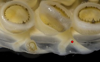
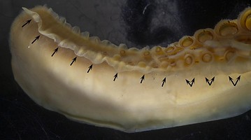
Figure. Sthenoteuthis oualaniensis. Left - Oral view of part of the ventral manus. Club tissue macerated making the trabeculae easier to see. One of the marginal suckers has been removed; the red dot represents the position and diameter of the cut neck of the sucker. Note that there are two trabeculae arising from each lateral sucker-stalk. This is characteristic only for the manus and only in ommastrephids (Abraliopsis spp. have something similar on some arms). Right - Oral-ventral view of the proximal club showing the ventral protective membrane merging into the ventral margin of the stalk and the double trabeculae (double arrows) of the former being replaced by single trabeculae (single arrows) on the carpus and stalk.
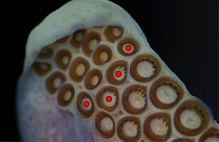
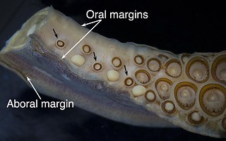
Figure. Sthenoteuthis oualaniensis, oral views of the proximal club. Left - Terminal pad. The red dots identify the terminal suckers of the dactylus which bear teeth on their inner rings in contrast to those of the terminal pad that have smooth inner rings. Right - Carpus. The carpus here is composed of three smooth-ringed suckers (black arrows) each with an adjacent, large, round, carpal-knob and located along the dorsal edge of the club. All other suckers in the region have toothed inner rings.
II. Internal structure: Skin, some suckers removed.
- Dorsal view: The lateral sucker-stalks are greatly enlarged on the mid-manus in spite of the relative small size of the suckers they carry; at the dactylus the sucker stalks decrease abruptly in size as does the large, trabeculate protective membrane.
- Ventral view: This opposite view shows the enlarged sucker-stalks of the mid-manus decreasing in a uniform manner to the tip of the dactylus along with the trabeculate protective membrane.
- The keel and the aboral margin of the stalk are continuous, that is they are both part of the same muscular structure.
- The proximal end of the keel is distinguished from the aboral margin by a by a low point in the structure near mid-manus (see yellow arrows below) where the keel has changed to a more dorsal position.
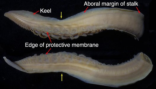
Figure. S. oualaniensis, 142 mm ML. Dorsal (top) and ventral views (bottom) of the clubs. The keel incorrectly seems to be in an aboral rather than a dorsal position due to the twist of the dactlyus (a fixation artifact - compare with photos of the same clubs in part I above). Note the two trabeculae per each marginal sucker-stalk, as seen above. Yellow arrows - Mark the low point in the continuum between the keel and the aboral margin of the stalk.
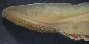
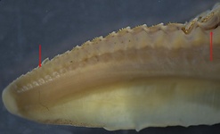
Figure. Photomicrographs of the dactylus, same club. Left - Dorsal view. Note the abrupt change in the depth of the membrane from the manus (right-arrow) to the dactylus (left-arrow) where the membrane is reduced to a low, rounded margin. Right - ventral view. Note the gradual change in the membrane from the manus (right arrow indicates the last sucker of the manus) toward the end of the dactylus (compare with "bottom" image in previous figure).
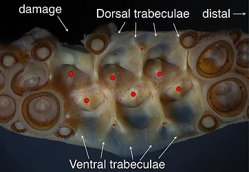
Figure. S. oualaniensis. Oral view of the distal manus with some suckers removed showing the complex arrangement of surface muscles. Red dots - Mark where sucker stalks were cut.
III. Internal structure: Cross-section of the mid-manus
- Organization: Manus somewhat bilaterally symmetrical with long stalks to lateral suckers and short stalks to medial suckers (irrespective of sucker sizes). (See "Basal clubs: Introduction" for comments on use of "bilateral symmetry.") Oral surface of manus basically flat when in relaxed state.
- Trabeculate protective membranes: Dorsal and ventral membranes large and equally developed.
- Core shape: Circular.
- Keel: Attached slightly dorsal to mid-aboral side of club core.
- Attachment of sucker stalks and trabeculae: Sucker stalks attach mostly to club core with some attachment to other sucker stalks; trabeculae attach to both the sucker stalks and club core. The complex musculature of the club is poorly understood.
- Open canals: None.
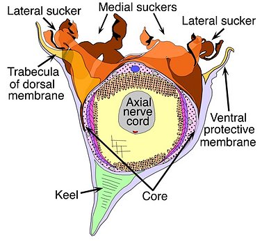
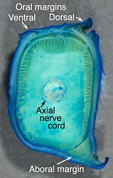
Figure. Left - Ommastrephes bartramii, 120 mm ML, cross-section through the manus showing the basic structure of the club core, and the attachment of the keel, sucker stalks and trabeculae of the protective membrane. Most of the core outside the axial nerve cord is composed of muscles. Dots on drawing areas indicate primarily longitudinal muscles. Other colors and fill-lines indicate more complex musculature. The red dot on the aboral side of the nerve cord is the major artery of the club; the blue dot oral of the nerve cord is the major vein of the club. Note that the stalks of the small, lateral suckers are longer than those of the large, medial suckers. The drawing, by R. Young, is a compilation taken from histological cross-sections. Right - Sthenoteuthis oualaniensis, xx mm ML. Cross-sectional cut with a scalpel of the tentacular stalk just proximal of the club, for comparison with the club.
- Organization: Strong asymmetry with very long stalks for the ventrolateral suckers grading to small stalks for dorsolateral suckers. Oral surface of dactylus basically flat, when in relaxed state, and in same plane with that of manus.
- Trabeculate protective membranes: Ventral trabecula and membrane well developed; dorsal counterparts greatly reduced.
- Core shape: Circular, tilted ventrally.
- Keel: Large; attached to dorsal side of core; oral surface (when relaxed) probably parallel to that of combined suckers.
- Attachment of sucker stalks and trabeculae: Uncertain.
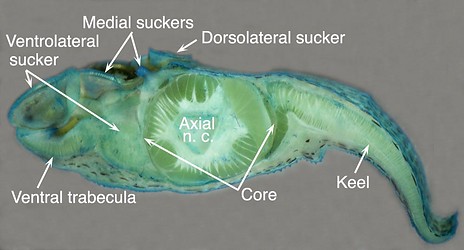
Figure. Sthenoteuthis oualaniensis. Transverse cut with scalpel showing differences in sucker-stalk and trabecula sizes between dorsal and ventral sides of the dactylus. The dorsomedial sucker lost the sucker and shows the sucker-stalk neck and short but broad base. The ventrolateral sucker has a very large base. Note the large ventral trabecula of the ventral protective membrane and the virtual absence of a dorsal trabeculate protective membrane.
General comments:
We assume that the carpus has secondarily been lost in the other ommastrephid subfamilies which have larger clubs and shorter tentacular stalks (e.g., see the drawing of the arm crown in Martilia hyadesi here), which would probably make a make a carpal locking-appartus less necessary. We consider (1) the presence of a keel continuous, in position and structure, with the aboral margin of the tentacular stalk and (2) the presence of trabeculae in the oral margins of the stalk to be primitive (i.e., plesiomorphic in decapodiforms) features as they both are characteristic of arms.


