


Brachioteuthidae
Marek Lipinski and Richard E. Young


This tree diagram shows the relationships between several groups of organisms.
The root of the current tree connects the organisms featured in this tree to their containing group and the rest of the Tree of Life. The basal branching point in the tree represents the ancestor of the other groups in the tree. This ancestor diversified over time into several descendent subgroups, which are represented as internal nodes and terminal taxa to the right.

You can click on the root to travel down the Tree of Life all the way to the root of all Life, and you can click on the names of descendent subgroups to travel up the Tree of Life all the way to individual species.
For more information on ToL tree formatting, please see Interpreting the Tree or Classification. To learn more about phylogenetic trees, please visit our Phylogenetic Biology pages.
close boxIntroduction
Brachioteuthids are small to medium sized squids (ca. 15 cm ML). The mantle is muscular but generally rather thin. The tentacular clubs are unusual: The dactylus is normal (four sucker series) but the proximal part of the manus is greatly expanded and carries numerous small suckers on long stalks. Another unusual feature is the compact digestive gland that is located well posterior to the cephalic cartilage in the mantle cavity in young squid (see photographs of the paralarvae below).
Little is known about the biology of brachioteuthids although Roper and Vecchione (1996) describe an accumulation of Brachioteuthis beanii observed from a submersible near the ocean floor at a depth of about 800 m.
Brief diagnosis
An oegopsid with ...
- buccal connectives attaching to ventral borders of arms IV.
- many series of irregular suckers on proximal manus; distal manus and dactylus usually with four regular series.
- paralarvae having elongate, extensile necks.
Characteristics
- Arms
- Arm suckers in two series.
- Buccal-crown connectives attach to ventral borders of arms IV.
- Tentacles
- Proximal region of manus of club greatly expanded with small suckers on long stalks in numerous irregular series.
- Dactylus of club with suckers in three or four series. Click on an image to view larger version & data in a new window
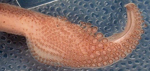
Figure. Oral view of tentacular club of Brachioteuthis sp., western North Atlantic. Photograph by M. Vecchione.
- Head
- Beaks: Descriptions of Brachioteuthis sp. A (probably B. beanii) can be found here: Lower beak, upper beak. See Sloarczykovia page for beaks of S. circumantarctica.
- Beaks: Descriptions of Brachioteuthis sp. A (probably B. beanii) can be found here: Lower beak, upper beak. See Sloarczykovia page for beaks of S. circumantarctica.
- Funnel
- Funnel-locking cartilage with straight groove.
- Funnel-locking cartilage with straight groove.
- Mantle
- Mantle muscular but thin.
- Mantle muscular but thin.
- Fins
- Fins short; anterior lobes free.
- Fins short; anterior lobes free.
- Photophores
- Single, large, ventral ocular photophore may or may not be present.
- Single, large, ventral ocular photophore may or may not be present.
- Viscera
- Compact digestive gland located well posterior to cephalic cartilage.
Nomenclature
A list of all nominal genera and species in the Brachioteuthidae can be found here. The list includes the current status and type species of all genera, and the current status, type repository and type locality of all species and all pertinent references.
Life history
Individual eggs are found in oblique plankton tows taken in the upper few hundred meters of the ocean. The eggs have a distinctive ellipsoidal shape. Presumably the eggs are spawned in either loose egg masses or strings that easily break apart or are spawned individually.
Members of the family have characteristic paralarvae. The paralarvae have long necks containing a fluid-filled sac that extends as a reservoir into the body (Young, et al., 1985). Contraction of the reservoir can greatly increase the length of the neck, thereby extending the head from the mantle. This trait is present at hatching.
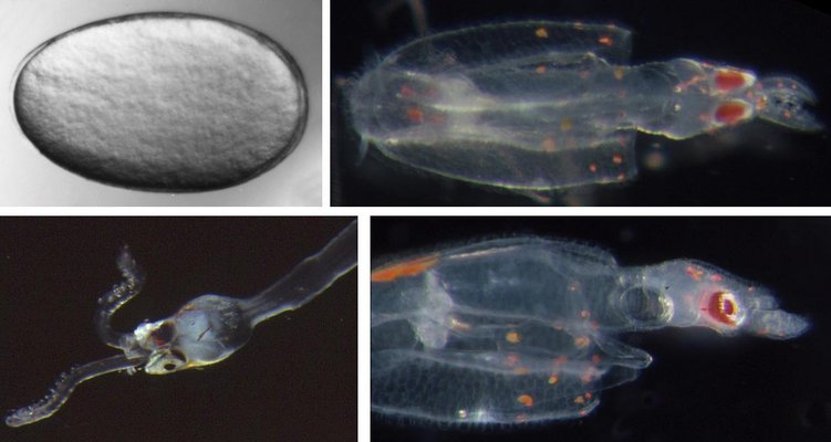
Figure. Ontogenetic stages of Brachioteuthis sp., Hawaiian waters. Top left - Egg. Top right - Dorsal view of hatchling, 2 mm ML, head retracted. Bottom left - Paralarva head and neck, 6 mm ML. Bottom right - Side view of the same hatchling, head retracted. Photographs by R. Young.
The photographs below were taken scuba diving at night off the leeward coast of Hawaii Island. This paralarva (ca. 6-10 mm ML) appears to have a maximum extension of its neck.Paralarvae are illustrated below showing change in form and chromatophore patterns.
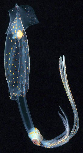
Brachioteuthis sp., same "older" paralarva as in
adjacent photograph. Side view. © 2015 Jeffrey Milisen

Figure. Brachioteuthis sp. paralarvae, composite photograph. Ventral views, taken in situ in oceanic
waters at night. Top - Younger paralarva (ca. 6-10 mm ML). Note the long neck and short arm crown.
Bottom - Older paralarva (ca. 15-20 mm ML). © Jeffrey Milisen
Paralarvae illustrated below show changes in form and chromatophore patterns with growth.
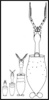
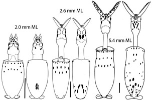

Figure. Ventral and dorsal views of four paralarval stages of Brachioteuthis sp., Hawaiian waters. Scale bar = 1 mm. The thumbnail at the left shows the relative sizes of the four stages - click it for a larger view. Drawings from Young et al. (1985).
The developing tentacular club is very unusual in that it has two separate areas in the distal club for sucker growth. Does this have broader phylogenetic implications?
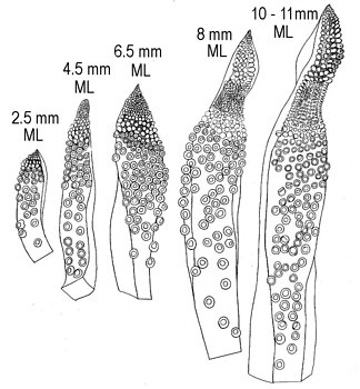
Figure. Brachioteuthis sp..Development of the paralarval tentacular club.Drawings from Degner, 1925.
References
Degner, E. 1925. Cephalopoda. Report on the Danish Oceanographical Expeditions 1908-10 to the Mediterranean and Adjacent Seas, 2(9):1-94.
Roper, C. F. E. and M. Vecchione (1996). In-situ observations on Brachioteuthis: paired behavior, possibly mating. Am. Malac. Bull., 13(12):55-60.
Young, R. E., R. F. Harman and K. M. Mangold (1985). The eggs and larvae of Brachioteuthis sp. (Cephalopoda: Teuthoidea) from Hawaiian waters. Vie Milieu, 35: 203-209.
Title Illustrations

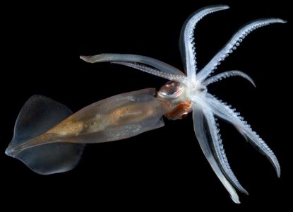
| Scientific Name | Brachioteuthis beanii |
|---|---|
| Location | Central North Atlantic |
| Comments | Note the large photophore on the ventral side of each eye. |
| Specimen Condition | Live Specimen |
| View | Ventral |
| Copyright |
©

|
About This Page
Sea Fisheries Institute, Cape Town, South Africa
University of Hawaii, Honolulu, HI, USA
Correspondence regarding this page should be directed to Marek Lipinski at
Page copyright © 2019 and
All Rights Reserved.
- Content changed 26 March 2019
Citing this page:
Lipinski, Marek and Richard E. Young. 2019. Brachioteuthidae . Version 26 March 2019. http://tolweb.org/Brachioteuthidae/19409/2019.03.26 in The Tree of Life Web Project, http://tolweb.org/

