


Ommastrephinae
Richard E. Young and Michael Vecchione
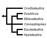


This tree diagram shows the relationships between several groups of organisms.
The root of the current tree connects the organisms featured in this tree to their containing group and the rest of the Tree of Life. The basal branching point in the tree represents the ancestor of the other groups in the tree. This ancestor diversified over time into several descendent subgroups, which are represented as internal nodes and terminal taxa to the right.

You can click on the root to travel down the Tree of Life all the way to the root of all Life, and you can click on the names of descendent subgroups to travel up the Tree of Life all the way to individual species.
For more information on ToL tree formatting, please see Interpreting the Tree or Classification. To learn more about phylogenetic trees, please visit our Phylogenetic Biology pages.
close boxIntroduction
Members of the Ommastrephinae are the most oceanic members of the Ommastrephidae and are commonly attracted to surface night-lights in the open ocean. Some species are fished commercially and one species, Ommastrephes bartramii, was taken in the North Pacific in quantities over 350,000 tons per year during the 1980s (Bower and Ichii, 2005). This subfamily contains the largest species (Doscidicus gigas) reaching a size of 120 cm ML and the smallest species (Hyaloteuthis pelagica) reaching a size of about 9 cm ML (Nesis, 1982/87). A wide variety of luminous organs are present on the mantle, head, arms, viscera and eyes but vary with species.
Brief diagnosis:
Ommastrephids with ...
- photophores.
Characteristics
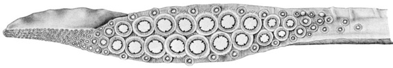
- Arms
- Hectocotylus usually with distal ventral protective membrane expanded to form a flange (not expanded in Ommastrephes where it tapers toward tip).
- Hectocotylus possesses pores in some genera.
Click on an image to view larger version & data in a new window
Figure. Mostly oral views of hectocotylized left or right ventral arms of representatives of Ommastrephinae genera. A - Ommastrephes bartramii. B - Sthenoteuthis pteropus, right arm. C - Dosidicus gigas, right arm. D - Eucleoteuthis luminosa, left arm. E - Hyaloteuthis pelagica, distal part of right arm, simplified. F - Ornithoteuthis volatilis, distal 2/3 right arm; left - oral view; right - ventrolateral view. v - Ventral side of arm. Drawings from Roeleveld (1988). Arrows point to pores.
- Tentacles
- Suckers of dactylus of tentacular club in four series.
- Carpal locking-apparatus usually present with one to several smooth-ringed suckers and corresponding knobs (absent in Ornithoteuthis).
- Head
- Funnel groove usually with anterior foveola and side pockets (both absent in Ornithoteuthis). Click on an image to view larger version & data in a new window
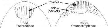
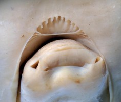
Figure. Left - Ventral view of the funnel groove comparing modifications in two of the subfamilies. Drawing modified from Roper, et al. (1985). Right - Oral-oblique view of the funnel (with funnel valve protruding) and funnel groove of Sthenoteuthis oualaniensis showing foveola and side pockets. Photograph by R. Young.
- Funnel groove usually with anterior foveola and side pockets (both absent in Ornithoteuthis).
- Photophores
- Photophores present in all species.
- Subcutaneous photophores present in all species except those of Ornithoteuthis.
Table. Comparison of genera of OmmastrephinaeClick on an image to view larger version & data in a new window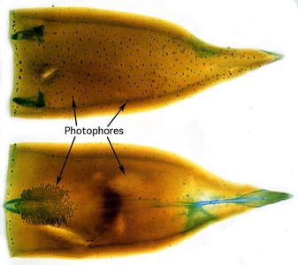
Figure. Ventral (top) and dorsal (bottom) views of the mantle of Sthenoteuthis oualaniensis that has been cleared and the photophores (and gladius, funnel locking-apparatus) stained with alcian blue stain. The numerous small, dark dots are subcutaneous photophores embedded in the mantle muscle. The lower, left arrow points to a dense patch of several hundred photophores. Photograph modified from Kishimoto and Kohno (1992).
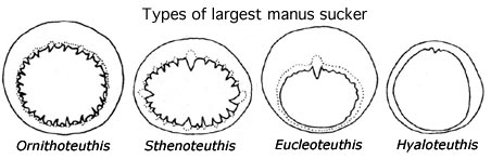
Largest manus sucker Visceral photophores Enlarged subcutaneous photophores Hectocotyllus with lateral pores Arm tips Ornithoteuthis Toothed, no enlarged teeth One or two round organs, streak None Yes Normal Dosidicus Toothed, enlarged tooth in each quadrant Two round organs, no streaks None Yes Attenuate at >35 cm ML, with 200-500 small suckers
Ommastrephes Toothed, enlarged tooth in each quadrant
None None No Normal
Sthenoteuthis Toothed, enlarged tooth in each quadrant
Two round organs, no streaks None Yes; No in early maturing form Normal
Eucleoteuthis Smooth except one tooth One round organ, no streaks Pads and streaks No Normal
Hyaloteuthis Smooth, sometimes one tooth One round organ, no streaks 19 circular pads No Normal
- Photophores present in all species.
References
Kishimoto, H. and H. Kohno. 1992. Development of the luminous organ in the purpleback flying squid, Sthenoteuthis oualaniensis, as shown by Alcian Blue stain techniques. Bull. Inst. Oceanic Res. & Develop., Tokai Univ, 13: 71-83.
Nesis, K. N. 1982. Abridged key to the cephalopod mollusks of the world's ocean. 385,ii pp. Light and Food Industry Publishing House, Moscow. (In Russian.). Translated into English by B. S. Levitov, ed. by L. A. Burgess (1987), Cephalopods of the world. T. F. H. Publications, Neptune City, NJ, 351pp.
Roeleveld, M. A. 1988. Generic interrelationships within the Ommastrephidae (Cephalopoda). P.277-314. In: M. R. Clarke and E. R. Trueman (eds.). The Mollusca. Vol. 12. Paleontology and Neontology of Cephalopods. Academic Press, N.Y., 355pp.
Roper, C. F. E., M. J. Sweeney, And C. E. Nauen. 1985. FAO species catalogue. Vol. 3. Cephalopods of the World. An annotated and illustrated catalogue of species of interest to fisheries. FAO Fish. Synop., (125)3:277 pp.
Title Illustrations
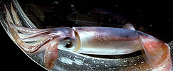
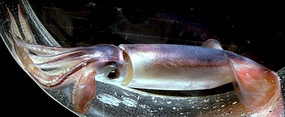
| Scientific Name | Sthenoteuthis oualaniensis |
|---|---|
| Location | Hawaiian waters |
| Specimen Condition | Live Specimen |
| View | Side |
| Size | Estimate of 18-20 cm ML |
| Copyright |
© 1996 Richard E. Young

|
About This Page
Richard E. Young
University of Hawaii, Honolulu, HI, USA
National Museum of Natural History, Washington, D. C. , USA
Page copyright © 2006 Richard E. Young and
- First online 06 April 2007
- Content changed 06 April 2007
Citing this page:
Young, Richard E. and Vecchione, Michael. 2007. Ommastrephinae . Version 06 April 2007 (under construction). http://tolweb.org/Ommastrephinae/19941/2007.04.06 in The Tree of Life Web Project, http://tolweb.org/

